Why Your Brain's Blood Supply Matters So Much in GBM
The human brain relies on a tightly controlled blood supply. A specialized wall called the blood-brain barrier (BBB) lines every blood vessel in the brain. It acts as a filter, keeping harmful molecules out while letting nutrients in. In a healthy brain, this barrier is nearly impenetrable.
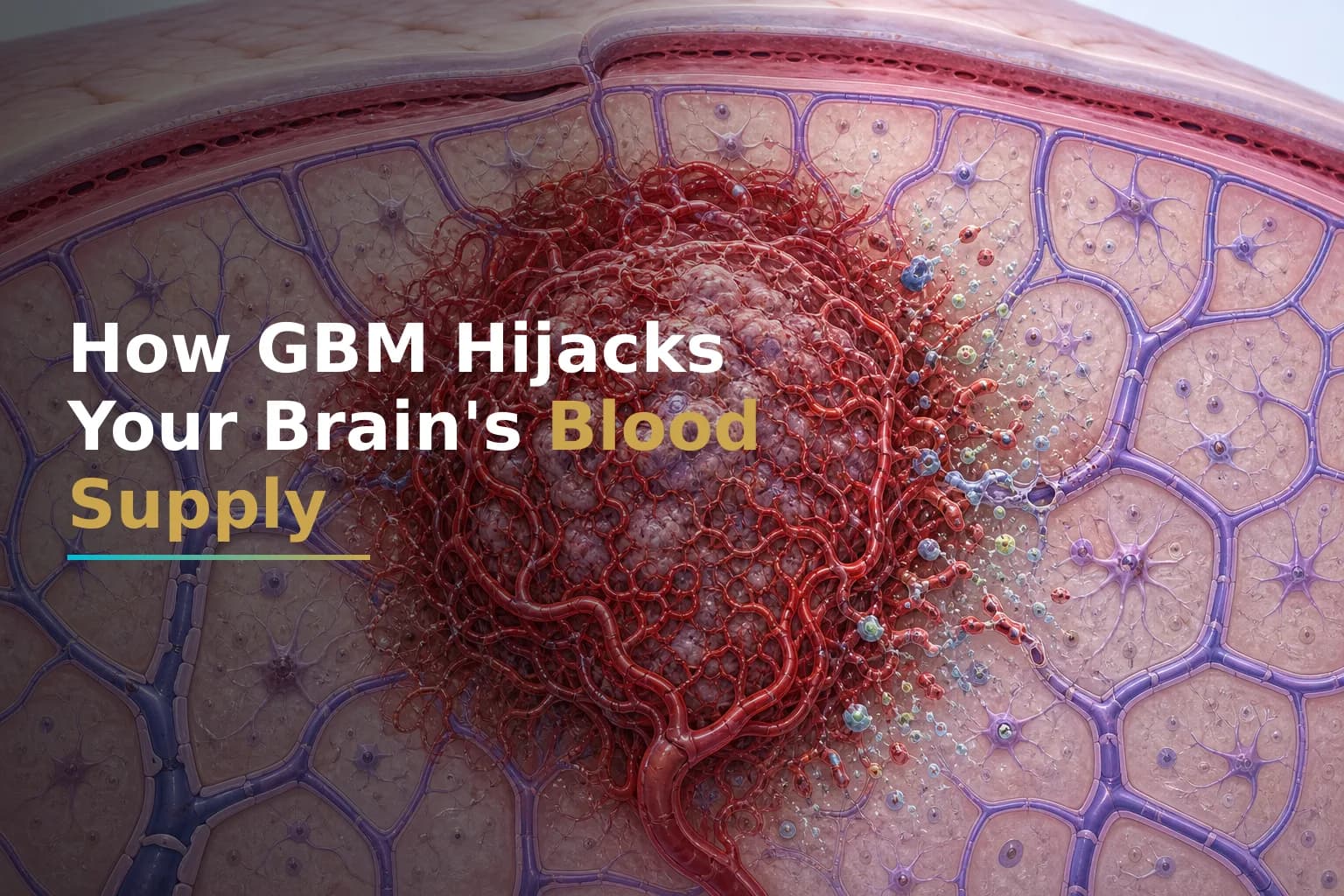
Glioblastoma (GBM) systematically dismantles that barrier. The tumor doesn't just grow — it engineers its own blood supply. It sends chemical signals that force nearby blood vessels to sprout new, abnormal branches, then weakens those vessels so they leak. The result is a chaotic vascular environment that feeds the tumor, floods surrounding brain tissue with fluid, and creates serious obstacles for therapy.
Understanding how this happens can help you make sense of your MRI scans, your symptoms, your steroid prescription, and why certain treatments exist. It also helps you ask sharper questions about emerging and integrative strategies your team may be considering.
What Is Angiogenesis — and Why Does GBM Need It?
Angiogenesis means the growth of new blood vessels from existing ones. Normally, it is tightly regulated — it supports wound healing and fetal development, then shuts off. Cancer cells hijack it for continuous use.
Research published in PMC describes glioblastoma as one of the most highly vascularized tumors known. One unique characteristic of glioblastoma is its highly vascularized nature, enabling the tumor to grow and invade surrounding brain tissue — a process known as angiogenesis, which is critical for the growth of glioblastoma cells.
As GBM grows, cells at the tumor core outpace their blood supply. Oxygen levels drop. This state — called hypoxia — triggers an emergency signaling cascade inside tumor cells.
Hypoxia in glioma tissue stimulates the expression of the transcription factor HIF-1α, which triggers the production of excess quantities of pro-angiogenic growth factors. One of the most pivotal of these is vascular endothelial growth factor A (VEGF-A).
VEGF-A works like a chemical distress signal. It tells nearby blood vessel cells to multiply and grow toward the tumor. The new vessels that form are structurally abnormal — disorganized, irregular in diameter, and poorly coated with the support cells (pericytes) that normally keep vessels stable. The result is a leaky, dysfunctional vascular network that serves the tumor while damaging surrounding brain tissue.
How GBM Breaks Down the Blood-Brain Barrier
The BBB is made up of several interacting components. The BBB is comprised of endothelial cells, pericytes, and astrocytes, forming a neurovascular unit that tightly regulates the transfer of ions and molecules between the blood and the brain.
GBM disrupts this unit through several mechanisms at once. Glioblastoma is distinguished by profound vascular abnormalities, including structurally disorganized vessels, reduced pericyte coverage, and compromised tight junctions. Elevated levels of VEGF drive aberrant angiogenesis, resulting in increased vascular permeability and focal BBB breakdown.
VEGF also attacks the molecular bonds that hold the BBB together. VEGF destabilizes tight junctions by promoting post-translational modification and mislocalization of junctional proteins such as occludin and ZO-1, resulting in increased paracellular permeability. In plain terms, the chemical bonds that seal the vessel wall are loosened, and fluid leaks through.
Other molecules add to the damage. The increased expression of molecules like metalloproteinases (MMP2-MMP9) and VEGF induces a breakdown of the BBB, especially the tight junctions of the endothelium, and enhances the penetration of tumoral cells.
Tumor-associated immune cells called macrophages also contribute. They release additional VEGF, angiopoietin-2, and MMP-9 — all potent disruptors of BBB integrity — helping sustain the cycle of vascular dysfunction as the tumor grows.
What This Looks Like on Your MRI
The vascular disruption caused by GBM directly affects how your tumor appears on imaging and how those images are interpreted.
Pixel intensity enhancement on the T1 gadolinium image corresponds to the contrast agent leaking from the tumor-induced neovasculature, while hyperintensity on the T2/FLAIR images corresponds with edema and infiltrated tumor cells.
The bright ring you may see on a contrast MRI is not the whole tumor — it marks where the BBB has broken down enough to let contrast dye leak in. But the picture is more complicated. Because of this leaky BBB, many researchers suggested that the BBB is no longer limiting drug delivery in GBM. However, there is increasing evidence that heterogeneous disruption of the BBB in GBM does not allow homogeneous and effective drug concentrations within tumor tissue — some portions of a malignant tumor may be protected by an undamaged BBB, such as the non-enhancing regions on MRI.
This means tumor cells hiding in non-enhancing regions — areas that look normal on an MRI — may still be shielded from chemotherapy. It is one reason GBM is so difficult to treat systemically.
For more on how your molecular profile interacts with these treatment barriers, see our article on Understanding Your GBM Molecular Profile: IDH, MGMT, EGFR & Why They Matter.
Edema: The Flooding That Causes Many of Your Symptoms
When the BBB breaks down and vessels leak, fluid builds up in the brain tissue around the tumor. This is called vasogenic edema. It is separate from the tumor itself, but it drives many of the neurological symptoms patients experience: headaches, fatigue, cognitive difficulties, weakness, and seizures.
VEGF is confirmed to be abnormally elevated in the pathogenesis of GBM, causing BBB pathological disruption, which further allows the leakage of neurotoxic blood-derived molecules into the central nervous system, interfering with brain homeostasis and leading to poor patient outcomes.
The standard short-term treatment for this edema involves corticosteroids. Common steroids used to treat brain tumor edema include dexamethasone, hydrocortisone, and prednisone. Dexamethasone works by modulating tight junction proteins such as claudin-5, occludin, and ZO-1, helping to partially restore BBB integrity and reduce fluid buildup. It can reduce symptoms within 24 to 48 hours.
However, dexamethasone's relationship with GBM outcomes is complex and actively studied. Dexamethasone is frequently administered in brain tumor patients for symptomatic relief. However, an increasing number of publications suggests that dexamethasone may lead to worse outcomes in patients with glioblastoma. Research from PMC reports that dexamethasone significantly reduces overall and progression-free survival in glioblastoma patients, even when accounting for clinical status. Additionally, steroids are also linked to a multitude of adverse side effects that may affect the survival of GBM patients, such as major immunosuppression and metabolic changes like hyperglycemia.
Dexamethasone relieves symptoms but may carry long-term costs. That is why many neuro-oncologists aim to use the lowest effective dose and taper as quickly as safely possible. A clinical trial listed on ClinicalTrials.gov (NCT04266977) is actively studying whether restricting dexamethasone use improves outcomes in GBM patients.
Anti-Angiogenic Therapy: Targeting VEGF Directly
Because VEGF is so central to GBM's vascular strategy, researchers developed drugs designed to block it. The most widely used is bevacizumab, a monoclonal antibody that binds VEGF-A and prevents it from activating receptors on blood vessel cells.
A critical protein that facilitates new blood vessel formation is VEGF-A; bevacizumab, a medication that specifically targets VEGF-A, has been approved for treatment of recurrent glioblastoma.
Bevacizumab can meaningfully reduce edema and improve how patients feel in the short term. By normalizing leaky tumor vessels, it reduces fluid buildup and can lower the need for dexamethasone. Its track record on survival, though, is more complicated. Despite its theoretical potential, bevacizumab has failed to offer significant survival improvement. Furthermore, other agents with comparable mechanisms of action have also not been able to demonstrate favorable results.
GBM adapts. While anti-angiogenic treatment showed promise, agents like bevacizumab have ultimately failed to improve overall survival. This highlights the presence of compensatory angiogenic mechanisms that bypass VEGF inhibition, necessitating further investigation into resistance mechanisms and the development of more effective therapeutic strategies.
GBM tumors can switch to alternative pro-angiogenic signals — angiopoietins, bFGF, PDGF — when VEGF is blocked. This escape behavior is an active area of research. For a closer look at how bevacizumab is used in practice, see our article on Bevacizumab for Recurrent Glioblastoma: What It Actually Does, Who It Helps, and What to Expect.
Why Drug Delivery Remains So Difficult
The disrupted BBB in GBM creates a paradox. In some areas, the barrier is broken, so drugs can enter. In others, including the infiltrating edge of the tumor, the BBB remains largely intact, blocking drugs from reaching tumor cells that are spreading into normal brain tissue.
This inconsistency means that even when a drug works well in a lab, it may not reach enough of the tumor at useful concentrations in a real patient. Two zones can often be observed within the tumor volume — zone 1: disrupted tumor BBB in the tumor core imaged via MRI contrast leakage; zone 2: intact tumor BBB in the tumor rim area.
This is why researchers are studying multiple strategies to either open the BBB selectively or engineer drugs that can cross it more effectively — including nanoparticles, focused ultrasound, and novel peptide carriers. Some of these approaches are in early clinical trials and represent an active frontier in GBM research.
Tumor Treating Fields (TTFields) may also interact with this system. Research has found that TTFields appear to alter junctional proteins at the BBB, potentially allowing a reversible, localized disruption that could improve drug penetration. For more on how TTFields work, see our article on Tumor Treating Fields (TTFields) for Glioblastoma: How the Optune Device Works, Who Qualifies, and What to Expect During Treatment.
Where Integrative Strategies May Have a Role
Several integrative and metabolic approaches are being studied in the context of GBM vascular biology. None of these replace standard-of-care treatment — surgery, radiation, and temozolomide remain the foundation of GBM management. Some research suggests they may address overlapping biological mechanisms, however.
- Ketogenic diet: By lowering blood glucose and shifting the metabolic environment, a ketogenic diet may reduce the signaling conditions that promote tumor angiogenesis. The hypoxia-VEGF axis is closely tied to glucose and energy metabolism. Early studies are underway. For context, see our article on The Ketogenic Diet and Glioblastoma: What the Evidence Actually Says About Using Metabolic Therapy Alongside Standard Treatment.
- Anti-inflammatory compounds: Chronic inflammation in the tumor microenvironment fuels ongoing VEGF secretion and BBB disruption. Some integrative compounds — including curcumin — are being studied for potential effects on this inflammatory signaling, though clinical evidence in GBM remains limited and preliminary. See our overview of Integrative Treatments for Glioblastoma: Evidence-Based Complementary Therapies That May Help.
- Drug repurposing candidates: Drugs originally developed for other conditions — including statins and metformin — are being examined for effects on angiogenic pathways. Some may interfere with VEGF signaling or alter the metabolic conditions that drive hypoxia. These are discussed in detail in separate articles on this site and should only be considered in consultation with your oncologist.
The core principle across these approaches: GBM's vascular dysregulation is not a passive side effect of tumor growth. It is an active strategy the tumor uses to sustain itself. Finding ways to interfere with that strategy while keeping toxicity manageable is one of the central challenges in GBM research today.
What This Means for You as a Patient
Understanding GBM's vascular biology can help you in several practical areas of your care:
- Reading your MRI: The contrast-enhancing region reflects where the BBB is most disrupted. The FLAIR signal reflects edema extent. Neither perfectly captures the full tumor burden.
- Your steroid prescription: Dexamethasone is given to protect brain function from edema. Your team should have a plan to use the minimum effective dose and taper when it is safe to do so.
- Anti-angiogenic drugs: Bevacizumab may significantly reduce edema and improve quality of life at recurrence, even if its effect on overall survival is modest. For many patients, that is a meaningful benefit.
- Drug delivery challenges: If a treatment works in some patients but not others, the uneven BBB may be part of the reason. This is an active area of research.
- Integrative additions: Strategies that support metabolic health and reduce inflammatory signaling may complement standard-of-care treatment — but should always be discussed with your oncology team before starting.
When to Talk to Your Doctor
Talk to your neuro-oncologist if you experience new or worsening headaches, sudden changes in vision, weakness, difficulty speaking, or seizures — these may signal changes in edema or tumor activity. Ask your team whether your current steroid dose is being actively managed and whether a taper plan is in place. If you are interested in anti-angiogenic therapy, metabolic interventions, or participation in a clinical trial targeting GBM vasculature, raise those questions at your next appointment. Your oncologist can review your specific molecular profile and imaging to advise whether those options fit your situation.
This article is for general information and is not a substitute for medical advice. Always consult your oncologist or care team about your specific situation.