Table of contents
Introduction
Glioblastoma multiforme (GBM) is the most common and aggressive primary malignant brain tumor in adults, classified as a WHO Grade IV astrocytoma. Despite decades of research, median survival remains approximately 14–16 months with standard treatment consisting of maximal surgical resection, radiotherapy, and temozolomide chemotherapy. The 5-year survival rate is less than 5%.
What makes GBM so formidable is its genetic and molecular heterogeneity. Even within a single tumor, different regions can harbor distinct genetic alterations, and the subclones respond differently to treatment. This intratumoral heterogeneity is a primary driver of therapeutic resistance and recurrence.
Understanding the genetics of glioblastoma is no longer academic. It is what identifies druggable targets, predicts treatment response, stratifies patients for clinical trials, and shapes a real treatment plan. The Cancer Genome Atlas (TCGA) mapped the genomic alterations in GBM in detail: oncogene amplifications, tumor suppressor deletions, point mutations, gene fusions, and epigenetic modifications, all interacting.
This guide provides a thorough exploration of every major genetic alteration known to drive glioblastoma, organized by pathway and mechanism, to serve as an educational resource for clinicians, researchers, and informed patients alike.
The molecular biology of glioblastoma
Glioblastoma is driven by the dysregulation of a relatively small number of critical signaling pathways. The Cancer Genome Atlas (TCGA) identified three core pathways altered in the vast majority of GBM tumors:
Three core pathways altered in GBM
These pathways collectively control cell growth, survival, proliferation, and death. What is particularly striking about GBM is that most tumors harbor alterations in all three pathways simultaneously, creating a synergistic effect that drives uncontrolled tumor growth and extraordinary resistance to therapy.
RTK / RAS / PI3K signaling
This pathway governs how cells respond to growth factors. When altered, it provides continuous pro-growth and pro-survival signals, even in the absence of external stimuli. Alterations include receptor amplification (EGFR, PDGFRA), activating mutations, and loss of negative regulators (NF1, PTEN).
p53 pathway
The p53 pathway acts as a critical checkpoint, halting cell division when DNA damage is detected and triggering apoptosis if repair is not possible. Loss of p53 function through TP53 mutations, MDM2/MDM4 amplification, or CDKN2A deletion allows damaged cells to continue dividing, accumulating further mutations.
RB cell cycle pathway
The retinoblastoma (RB) pathway regulates the G1/S transition in the cell cycle. When disrupted through RB1 loss, CDK4/6 amplification, or CDKN2A/B deletion, cells bypass this critical checkpoint and enter an unrestrained proliferative state.
| Pathway | Frequency in GBM | Key Genes | Biological Effect |
|---|---|---|---|
| RTK/RAS/PI3K | ~88% | EGFR, PDGFRA, PTEN, NF1, PIK3CA | Growth & survival signaling |
| p53 | ~87% | TP53, MDM2, MDM4, CDKN2A | Evasion of apoptosis |
| RB | ~78% | RB1, CDK4, CDK6, CDKN2A/B | Uncontrolled cell division |
Receptor tyrosine kinase (RTK) pathway alterations
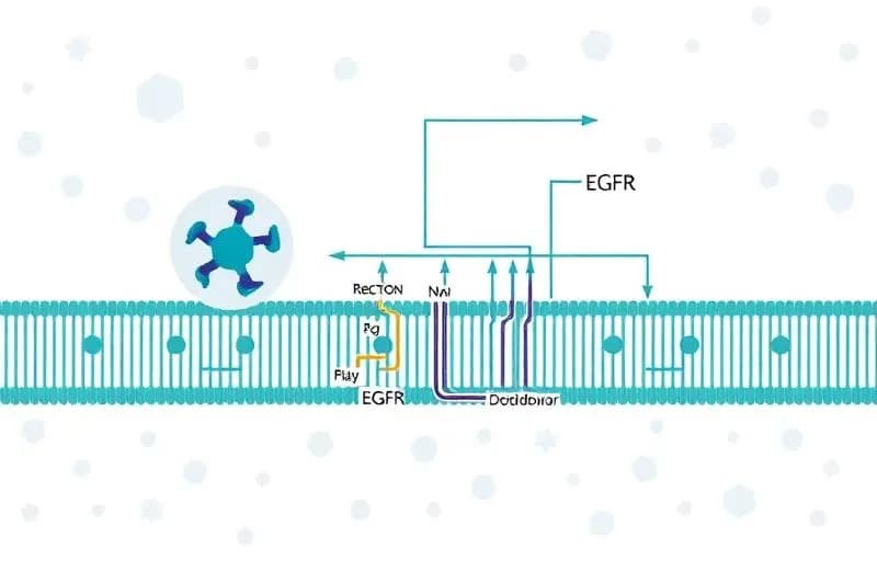
RTK signaling pathway
Receptor tyrosine kinases like EGFR sit on the cell membrane and relay growth signals into the cell. When amplified or mutated, they drive uncontrolled tumor growth.
Receptor tyrosine kinases (RTKs) are transmembrane proteins that relay extracellular growth signals into the cell. In GBM, these receptors are frequently amplified, mutated, or overexpressed, leading to constitutive activation of downstream growth and survival pathways. RTK alterations are the most common class of genetic changes in glioblastoma.
EGFR amplification & EGFRvIII
Epidermal Growth Factor Receptor (EGFR) is the most frequently amplified gene in GBM, occurring in approximately 40–50% of primary glioblastomas. EGFR amplification leads to receptor overexpression and constitutive activation of the RAS/MAPK and PI3K/AKT signaling cascades, driving tumor cell proliferation, survival, and invasion.
A particularly important variant is EGFRvIII (variant III), a truncated form of EGFR that results from an in-frame deletion of exons 2–7. EGFRvIII is constitutively active without requiring ligand binding and is found in approximately 25–30% of EGFR-amplified GBMs. It is a tumor-specific antigen not expressed in normal tissue, making it an attractive therapeutic target for vaccines and antibody-drug conjugates.
PDGFRA amplification
Platelet-Derived Growth Factor Receptor Alpha (PDGFRA) is amplified in approximately 10–15% of GBMs, particularly in the proneural molecular subtype. PDGFRA amplification creates autocrine signaling loops that promote gliomagenesis. The PDGFRA Δ8,9 deletion mutant is analogous to EGFRvIII and is constitutively active.
MET amplification
MET (hepatocyte growth factor receptor) amplification occurs in approximately 4% of primary GBMs but is significantly enriched in recurrent tumors, where it is a key resistance mechanism to EGFR-targeted therapies. MET activation promotes tumor invasion, angiogenesis, and stem cell maintenance.
FGFR mutations
Fibroblast Growth Factor Receptor (FGFR) alterations, including FGFR1, FGFR2, and FGFR3 mutations and fusions, occur in a small subset of GBM. FGFR3-TACC3 fusions are particularly notable as they represent actionable targets for FGFR inhibitors currently in clinical trials.
VEGFA amplification
Vascular Endothelial Growth Factor A (VEGFA) is not a receptor tyrosine kinase itself, but is a critical ligand that drives tumor angiogenesis through VEGFR signaling. GBM is among the most vascularized of all solid tumors. VEGFA overexpression drives the formation of the abnormal, leaky vasculature characteristic of GBM and contributes to peritumoral edema. Bevacizumab, an anti-VEGF antibody, is used in recurrent GBM management.
NF1 mutations
Neurofibromin 1 (NF1) is a tumor suppressor that negatively regulates RAS signaling. NF1 loss-of-function mutations, occurring in approximately 10% of GBMs, lead to constitutive RAS activation. NF1 mutations define the mesenchymal molecular subtype and are associated with a more aggressive, immune-infiltrated tumor microenvironment.
| Gene | Alteration Type | Frequency | Biological Effect |
|---|---|---|---|
| EGFR | Amplification / EGFRvIII | 40–50% | Constitutive growth signaling |
| PDGFRA | Amplification | 10–15% | Autocrine growth loops |
| MET | Amplification | ~4% | Invasion, therapy resistance |
| FGFR1/2/3 | Mutation / Fusion | ~3% | Growth signaling |
| VEGFA | Overexpression | High | Angiogenesis |
| NF1 | Loss-of-function | ~10% | Constitutive RAS activation |
PI3K / AKT / mTOR pathway
The PI3K/AKT/mTOR pathway is one of the most critical intracellular signaling cascades in GBM. It regulates cell survival, metabolism, growth, and protein synthesis. This pathway is activated downstream of RTKs and is the most frequently altered intracellular signaling pathway in glioblastoma.
PI3K / AKT / mTOR signaling cascade
PTEN loss
Phosphatase and Tensin Homolog (PTEN) is a critical tumor suppressor that negatively regulates PI3K signaling by dephosphorylating PIP3. PTEN is lost or mutated in approximately 30–40% of GBMs through homozygous deletion, mutation, or promoter methylation. PTEN loss leads to constitutive AKT activation and is associated with aggressive tumor behavior and resistance to multiple therapies.
PIK3CA and PIK3R1 mutations
PIK3CA encodes the catalytic subunit (p110α) of PI3K. Activating mutations occur in approximately 10–15% of GBMs, leading to constitutive PI3K activity. PIK3R1 encodes the regulatory subunit (p85α); mutations in this gene (found in ~8% of GBMs) disrupt its ability to inhibit the catalytic subunit, similarly resulting in pathway hyperactivation.
AKT and mTOR activation
AKT is a serine/threonine kinase at the central node of PI3K signaling. When activated, AKT phosphorylates downstream targets that promote cell survival (by inhibiting pro-apoptotic factors), metabolism, and growth. AKT directly activates mTOR (mechanistic Target of Rapamycin), a master regulator of protein synthesis and cell growth that integrates signals from nutrients, growth factors, and energy status.
TSC1/TSC2 alterations
Tuberous Sclerosis Complex proteins TSC1 (hamartin) and TSC2 (tuberin) form a complex that negatively regulates mTOR signaling. Although mutations in TSC1/TSC2 are rare in GBM, their functional loss through pathway-level dysregulation contributes to mTOR hyperactivation. These proteins serve as a critical brake on mTOR, and their dysfunction removes an important layer of growth control.
| Gene | Role | Alteration | Consequence |
|---|---|---|---|
| PTEN | Tumor suppressor | Deletion / Mutation | Constitutive AKT activation |
| PIK3CA | PI3K catalytic subunit | Activating mutation | PI3K hyperactivation |
| PIK3R1 | PI3K regulatory subunit | Loss-of-function | Loss of PI3K regulation |
| AKT | Kinase | Activation | Survival and growth |
| mTOR | Growth regulator | Hyperactivation | Protein synthesis, metabolism |
| TSC1/TSC2 | mTOR inhibitor | Functional loss | Unrestrained mTOR |
The p53 tumor suppressor pathway
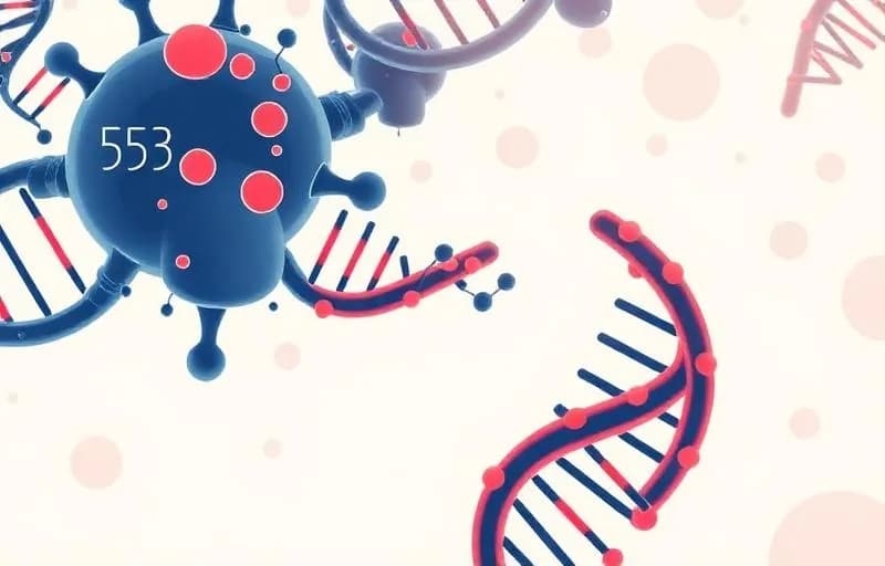
The guardian of the genome
p53 acts as a critical checkpoint - detecting DNA damage and triggering repair or cell death. When lost in GBM, damaged cells survive and accumulate further mutations.
The p53 pathway is one of the most important tumor suppressor networks in human biology. TP53, often called the "guardian of the genome," encodes the p53 transcription factor that responds to DNA damage, oncogene activation, and cellular stress by inducing cell cycle arrest, DNA repair, senescence, or apoptosis. In GBM, the p53 pathway is disrupted in approximately 87% of tumors.
p53 pathway in normal cells vs GBM
TP53 mutations
TP53 mutations occur in approximately 28–35% of GBMs, predominantly as missense mutations in the DNA-binding domain. These mutations are particularly prevalent in secondary GBMs that evolve from lower-grade gliomas. Mutant p53 loses its tumor suppressor function, and some mutations gain oncogenic properties (gain-of-function mutations) that actively promote invasion, genomic instability, and therapy resistance.
MDM2 and MDM4 amplification
MDM2 is an E3 ubiquitin ligase that targets p53 for proteasomal degradation. MDM2 amplification, found in approximately 8–10% of GBMs, effectively silences p53 function even when the TP53 gene itself is wild-type. MDM4 (MDMX) similarly inhibits p53 transcriptional activity and is amplified in approximately 4–7% of GBMs. Together, MDM2 and MDM4 amplification represent an alternative mechanism of p53 pathway inactivation.
CDKN2A deletion
CDKN2A encodes two distinct tumor suppressors through alternative reading frames: p14ARF (which stabilizes p53 by inhibiting MDM2) and p16INK4a (which inhibits CDK4/6 in the RB pathway). Homozygous deletion of CDKN2A, found in approximately 50–60% of GBMs, simultaneously disrupts both the p53 and RB pathways, making it one of the most consequential single genetic events in GBM.
RB cell cycle pathway
The retinoblastoma (RB) pathway is a critical regulator of the G1/S transition in the cell cycle. In normal cells, the RB protein acts as a molecular brake, preventing cells from entering S phase (DNA synthesis) until appropriate growth signals are received. In GBM, this pathway is disrupted in approximately 78% of tumors, releasing the brake on cell division.
RB pathway: cell cycle control
RB1 loss
RB1 loss through mutation or deletion occurs in approximately 8–12% of GBMs. When RB1 is lost, E2F transcription factors are constitutively active, driving continuous expression of genes required for DNA replication and cell division. RB1 loss is mutually exclusive with CDK4 amplification in most cases, as both achieve the same functional outcome.
CDK4 and CDK6 amplification
CDK4 amplification occurs in approximately 14–18% of GBMs and is a hallmark of the classical molecular subtype. CDK4, in complex with Cyclin D, phosphorylates and inactivates RB, mimicking the effect of RB1 loss. CDK6 amplification is less common but functionally similar. These amplifications are attractive therapeutic targets for CDK4/6 inhibitors (palbociclib, ribociclib, abemaciclib), although clinical results in GBM have been challenging.
Cyclin D alterations and CDKN2A/B deletion
Cyclin D1 (CCND1) and Cyclin D2 (CCND2) amplification provide increased binding partners for CDK4/6, enhancing RB phosphorylation. The CDKN2A and CDKN2B loci encode p16INK4a and p15INK4b, respectively, which are endogenous CDK4/6 inhibitors. Their homozygous co-deletion (found in ~50% of GBMs) removes a critical layer of cell cycle control.
Telomere maintenance mechanisms
Telomeres are protective caps at the ends of chromosomes that shorten with each cell division. In normal cells, progressive telomere shortening triggers replicative senescence or apoptosis-a powerful barrier against unlimited proliferation. Cancer cells must overcome this barrier through telomere maintenance mechanisms to achieve replicative immortality, one of the hallmarks of cancer.
TERT promoter mutations
Telomerase Reverse Transcriptase (TERT) promoter mutations are among the most common genetic alterations in primary GBM, occurring in approximately 70–80% of cases. Two hotspot mutations (C228T and C250T) create de novo binding sites for ETS transcription factors, driving TERT upregulation and telomerase reactivation. TERT promoter mutations are characteristic of primary (IDH-wildtype) GBM and are associated with older patient age.
ATRX and DAXX mutations
ATRX and DAXX form a chromatin remodeling complex that deposits the histone variant H3.3 at telomeric regions. Loss-of-function mutations in ATRX (occurring in ~7% of GBMs, predominantly IDH-mutant tumors) or DAXX activate the Alternative Lengthening of Telomeres (ALT) pathway, a recombination-based mechanism that maintains telomeres without telomerase. ALT-positive tumors have a distinctive phenotype and may respond differently to certain therapies.
| Mechanism | Gene | Frequency | GBM Subtype Association |
|---|---|---|---|
| Telomerase activation | TERT | 70–80% | IDH-wildtype (primary) |
| ALT pathway | ATRX | ~7% | IDH-mutant (secondary) |
| ALT pathway | DAXX | Rare | IDH-mutant |
IDH mutations and secondary glioblastoma
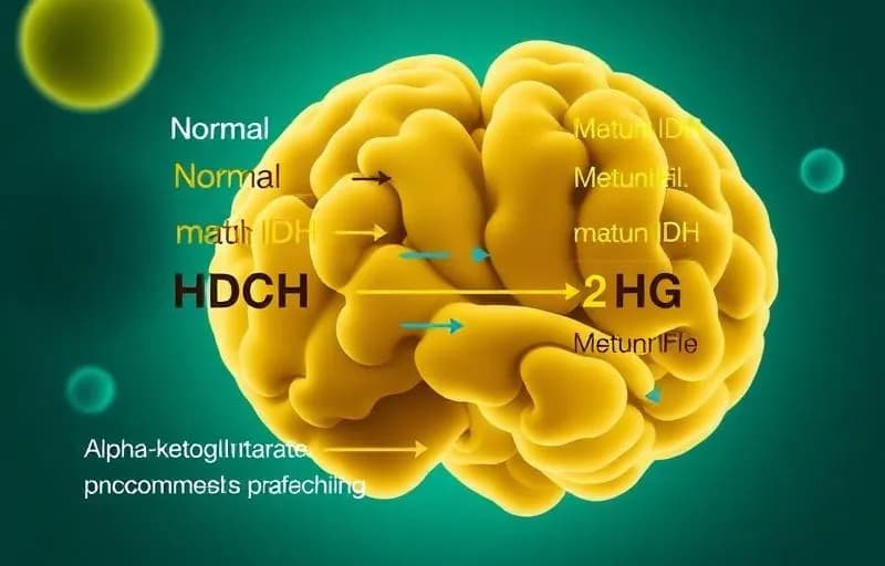
IDH mutation & metabolic reprogramming
Mutant IDH enzymes produce the oncometabolite 2-HG, which reprograms cell epigenetics and separates two biologically distinct diseases.
Isocitrate Dehydrogenase (IDH) mutations are one of the most significant molecular discoveries in glioma biology. They separate two biologically distinct diseases that were historically lumped together under the label "glioblastoma."
Primary vs secondary GBM
Primary (de novo) GBM accounts for approximately 90% of cases and arises without a clinically evident precursor lesion. These tumors are IDH-wildtype and are characterized by EGFR amplification, PTEN loss, TERT promoter mutations, and chromosome 7 gain/chromosome 10 loss. Secondary GBM evolves from lower-grade diffuse gliomas (WHO Grade II or III) over months to years and is characterized by IDH mutations, TP53 mutations, and ATRX loss.
Important note: Under the 2021 WHO Classification of CNS Tumors, IDH-mutant tumors are no longer classified as "glioblastoma" regardless of grade. They are now classified as "Astrocytoma, IDH-mutant, WHO Grade 4." The term "glioblastoma" is reserved exclusively for IDH-wildtype tumors. However, the biology of IDH mutations remains critically important for understanding glioma evolution.
IDH1 and IDH2 mutations
IDH1 R132H is the most common IDH mutation (>90% of IDH-mutant gliomas), occurring at a conserved arginine residue in the enzyme's active site. IDH2 R172 mutations are less common and functionally equivalent. These mutations are heterozygous and neomorphic-rather than simply losing function, the mutant enzyme gains a new activity: converting α-ketoglutarate to the oncometabolite 2-hydroxyglutarate (2-HG).
2-HG accumulation has broad metabolic and epigenetic consequences. It competitively inhibits α-ketoglutarate-dependent dioxygenases, including TET2 (involved in DNA demethylation) and Jumonji-domain histone demethylases. This produces the CpG island methylator phenotype (G-CIMP), widespread DNA and histone hypermethylation that reshapes gene expression programs.
| Feature | Primary GBM (IDH-wildtype) | Secondary (IDH-mutant Grade 4) |
|---|---|---|
| Frequency | ~90% | ~10% |
| Age of onset | Older (median ~62) | Younger (median ~44) |
| Precursor | De novo | Lower-grade glioma |
| Key mutations | EGFR, PTEN, TERT | IDH1/2, TP53, ATRX |
| Prognosis | Worse (~14 months) | Better (~31 months) |
| Epigenetics | G-CIMP negative | G-CIMP positive |
Epigenetic and chromatin regulation alterations
Beyond mutations in classical oncogenes and tumor suppressors, GBM frequently harbors alterations in genes that regulate chromatin structure and epigenetic marks. These mutations do not directly drive proliferation but they reshape gene expression programs, differentiation states, and genomic stability.
The SWI/SNF complex deserves special mention-it is a multi-subunit chromatin remodeling machine that uses ATP hydrolysis to reposition nucleosomes and regulate gene accessibility. Mutations in SWI/SNF subunits (ARID1A, ARID1B, SMARCA4, SMARCB1) are collectively found in approximately 10–15% of GBMs and represent a distinct mechanism of tumor suppression loss that is increasingly recognized across many cancer types.
DNA repair alterations
DNA repair pathways matter in GBM for two reasons. They shape genomic stability and tumor evolution, and they directly determine sensitivity to alkylating chemotherapy (temozolomide), the cornerstone of GBM treatment.
MGMT promoter methylation
O6-Methylguanine-DNA Methyltransferase (MGMT) is arguably the most clinically significant molecular biomarker in GBM. MGMT is a DNA repair enzyme that removes alkyl groups from the O6 position of guanine, directly counteracting the cytotoxic effect of temozolomide. When the MGMT promoter is methylated (found in approximately 35–45% of GBMs), the gene is silenced, and tumor cells cannot repair temozolomide-induced DNA damage-leading to significantly better treatment response and survival.
MGMT promoter methylation status is the strongest independent predictor of benefit from temozolomide and is routinely tested in clinical practice. Patients with methylated MGMT promoter have median survival of approximately 21–23 months compared to 12–15 months for those with unmethylated promoters.
Mismatch repair (MMR) deficiency
The mismatch repair system (MLH1, MSH2, MSH6, PMS2) corrects base-base mismatches and small insertion/deletion loops during DNA replication. MMR deficiency in GBM can be primary (rare) or acquired during temozolomide treatment. Temozolomide-induced hypermutation through MMR loss is a recognized resistance mechanism in recurrent GBM-the tumor develops thousands of new mutations, increasing heterogeneity and rendering it resistant to further alkylating therapy. MMR-deficient recurrent GBMs may, however, respond to immune checkpoint inhibitors due to their high tumor mutational burden.
| Gene | Function | Clinical Significance |
|---|---|---|
| MGMT | Alkyl group removal | Methylation = TMZ sensitivity, better prognosis |
| MSH6 | Mismatch repair | Loss = TMZ resistance, hypermutation |
| MSH2 | Mismatch repair | Deficiency = microsatellite instability |
| MLH1 | Mismatch repair | Loss may enable checkpoint immunotherapy |
| PMS2 | Mismatch repair | Contributes to MMR deficiency |
Gene fusions in glioblastoma
Gene fusions result from chromosomal rearrangements that join parts of two different genes, creating novel fusion proteins with oncogenic properties. While individually rare in GBM, gene fusions are critically important because many are actionable therapeutic targets with FDA-approved or investigational inhibitors.
FGFR3–TACC3 fusion
The FGFR3–TACC3 fusion is the most common gene fusion in GBM, occurring in approximately 3% of cases. This fusion retains the kinase domain of FGFR3 and the coiled-coil domain of TACC3, creating a constitutively active kinase that localizes to mitotic spindles and drives aneuploidy. It is targetable with FGFR inhibitors such as erdafitinib and futibatinib.
NTRK fusions
Neurotrophic Tyrosine Receptor Kinase (NTRK1, NTRK2, NTRK3) fusions are rare in adult GBM (<1%) but are enriched in pediatric high-grade gliomas. These fusions are highly actionable with FDA-approved TRK inhibitors larotrectinib and entrectinib, which have demonstrated durable responses in NTRK-fusion-positive tumors across histologies.
Other actionable fusions
| Fusion | Frequency | Approved/Investigational Drug | Status |
|---|---|---|---|
| FGFR3-TACC3 | ~3% | Erdafitinib, futibatinib | Clinical trials |
| NTRK1/2/3 | <1% | Larotrectinib, entrectinib | FDA-approved (agnostic) |
| ROS1 | Very rare | Crizotinib, entrectinib | FDA-approved (other cancers) |
| ALK | Very rare | Alectinib, lorlatinib | FDA-approved (other cancers) |
| MET | Rare | Capmatinib, tepotinib | Clinical trials in GBM |
Chromosomal abnormalities
Chromosomal gains & losses
GBM is characterized by large-scale chromosomal changes - chromosome 7 gain paired with chromosome 10 loss is the most consistent cytogenetic signature, affecting ~80% of cases.
Beyond individual gene-level alterations, GBM is characterized by large-scale chromosomal gains and losses that affect entire arms or chromosomes. These copy number alterations are among the most consistent features of GBM and affect the dosage of multiple oncogenes and tumor suppressors simultaneously.
Chromosome 7 gain and chromosome 10 loss
Combined gain of chromosome 7 and loss of chromosome 10 (+7/−10) is the most characteristic cytogenetic signature of IDH-wildtype GBM, found in approximately 80% of cases. Chromosome 7 harbors EGFR (7p11.2), MET (7q31.2), and other growth-promoting genes. Chromosome 10 harbors PTEN (10q23.3), the critical PI3K pathway tumor suppressor. The combined effect of EGFR gain and PTEN loss creates a powerful oncogenic synergy.
9p21 deletion (CDKN2A/B locus)
Homozygous deletion of the 9p21 locus, encompassing CDKN2A and CDKN2B, is found in approximately 50–60% of GBMs. As discussed in Sections 5 and 6, this single deletion simultaneously disrupts both the p53 pathway (via p14ARF) and the RB pathway (via p16INK4a and p15INK4b).
Other chromosomal changes
Molecular subtypes of glioblastoma
Gene expression profiling studies, led by the TCGA consortium, identified distinct molecular subtypes of GBM based on transcriptomic signatures. While the original classification described four subtypes, the neural subtype has been largely attributed to contamination by normal brain tissue, and current consensus recognizes three robust subtypes.
Classical subtype
The classical subtype is defined by high-level EGFR amplification and EGFRvIII expression, chromosome 7 gain, chromosome 10 loss, and CDKN2A homozygous deletion. TP53 mutations are notably absent. This subtype shows the strongest response to aggressive radiation and chemotherapy protocols and has a gene expression pattern resembling astrocytic lineage cells.
Mesenchymal subtype
The mesenchymal subtype is characterized by NF1 loss/mutation, high expression of mesenchymal markers (CHI3L1/YKL-40, MET, CD44), prominent immune infiltration, and necrosis. This subtype resembles a wound-healing or inflammatory state and is associated with the most aggressive clinical behavior but may respond to immunotherapy approaches due to its high immune content.
Proneural subtype
The proneural subtype is characterized by PDGFRA amplification, IDH1 mutations (in a subset), TP53 mutations, and gene expression patterns resembling oligodendrocyte progenitor cells. It tends to occur in younger patients and historically has had a slightly better prognosis, although it responds poorly to standard radiation/temozolomide therapy.
Neural subtype (deprecated)
The neural subtype was originally described as expressing neuron-associated genes but has since been largely attributed to contamination of tumor samples with normal brain tissue at the tumor margins. It is no longer considered a true GBM subtype in most current classifications.
| Subtype | Key Genetic Features | Gene Expression | Clinical Features |
|---|---|---|---|
| Classical | EGFR amp, +7/−10, CDKN2A del | Astrocytic | Best response to chemoradiation |
| Mesenchymal | NF1 loss, high immune markers | Mesenchymal/inflammatory | Most aggressive, immune-infiltrated |
| Proneural | PDGFRA amp, IDH1 mut, TP53 mut | OPC-like | Younger patients, poor TMZ response |
| Neural* | Mixed | Neuronal | *Likely contamination artifact |
Clinical significance: Subtype classification is not yet used for routine treatment decisions but is increasingly important in clinical trial design and may guide future personalized therapy approaches. Importantly, GBM subtypes are not static-tumors can shift between subtypes during treatment and recurrence, with a frequent transition toward the mesenchymal phenotype at recurrence.
Clinical implications of genetic alterations
The wealth of genomic data now available for GBM has increasingly important implications across the entire clinical management spectrum.
Prognosis
Several genetic biomarkers provide independent prognostic information. MGMT promoter methylation is the strongest predictor of overall survival with standard therapy. IDH mutation status separates tumor biology and survival expectations. CDKN2A homozygous deletion is an adverse prognostic factor, particularly in lower-grade gliomas progressing to high-grade disease. TERT promoter mutations combined with EGFR amplification define the most aggressive primary GBM phenotype.
Therapy selection
MGMT promoter methylation status increasingly guides treatment decisions, particularly in elderly patients where the balance between treatment benefit and toxicity is critical. Patients with unmethylated MGMT may benefit more from alternative approaches such as tumor-treating fields (TTFields) or clinical trial enrollment. Specific genetic alterations open doors to targeted therapies: FGFR fusions for FGFR inhibitors, NTRK fusions for TRK inhibitors, BRAF V600E mutations for BRAF/MEK inhibitor combinations, and potential CDK4/6 inhibitor applications for CDK4-amplified tumors.
Resistance mechanisms
Understanding genetic alterations is essential for anticipating and overcoming treatment resistance. Temozolomide resistance through MGMT expression or MMR deficiency, radiation resistance through enhanced DNA repair or stem cell phenotypes, and targeted therapy resistance through pathway redundancy (e.g., MET amplification as an escape from EGFR inhibition) all have genetic underpinnings.
Clinical trials and personalized medicine
Modern clinical trials increasingly use molecular stratification for patient selection. Basket trials enroll patients based on genetic alterations regardless of tumor histology, and umbrella trials test multiple therapies within GBM based on molecular profiles. Comprehensive genomic profiling through next-generation sequencing panels is becoming standard of care, identifying actionable alterations in approximately 20–30% of GBM patients.
Future directions in glioblastoma treatment
The future of precision oncology
Genomic profiling, immunotherapy, and targeted agents are turning GBM treatment away from a single template toward plans built around each tumor's biology.
The genomic understanding of GBM is opening therapeutic strategies that go beyond treating every patient with the same protocol.
Precision oncology
Comprehensive molecular profiling at diagnosis and recurrence, using technologies such as whole-genome sequencing, RNA sequencing, and single-cell analysis, is enabling truly personalized treatment plans. Molecular tumor boards integrate genomic data with clinical context to identify optimal therapeutic strategies for individual patients.
Immunotherapy
Despite the immunologically "cold" nature of most GBMs, several immunotherapy approaches show promise. CAR-T cell therapy targeting EGFRvIII, IL13Rα2, or GD2 has demonstrated responses in early clinical trials. Cancer vaccines (peptide-based and dendritic cell vaccines) are being refined based on tumor-specific neoantigens. Immune checkpoint inhibitors may benefit the subset of GBMs with high tumor mutational burden due to MMR deficiency.
Targeted therapy
Next-generation targeted agents with improved brain penetrance are in development for virtually every major altered pathway in GBM. Combination approaches targeting multiple pathways simultaneously aim to overcome the redundancy and resistance that have plagued single-agent strategies.
Metabolic targeting
GBM cells run on altered metabolism - aerobic glycolysis (the Warburg effect), glutamine addiction, and reprogrammed lipid metabolism - and each of those dependencies opens a therapeutic avenue. Approaches include ketogenic diets, 2-deoxy-D-glucose (), IDH inhibitors for IDH-mutant tumors, and mTOR-targeted metabolic interventions.
Genomic profiling and liquid biopsy
Circulating tumor DNA (ctDNA) detection in cerebrospinal fluid and blood is emerging as a minimally invasive approach to monitor treatment response, detect recurrence early, and track clonal evolution in real time. This technology promises to transform GBM management by enabling dynamic treatment adaptation based on evolving tumor genetics.
Knowledge is the first step
Understanding the genetics of glioblastoma helps patients, families, and clinicians make informed decisions about treatment and clinical trial participation.