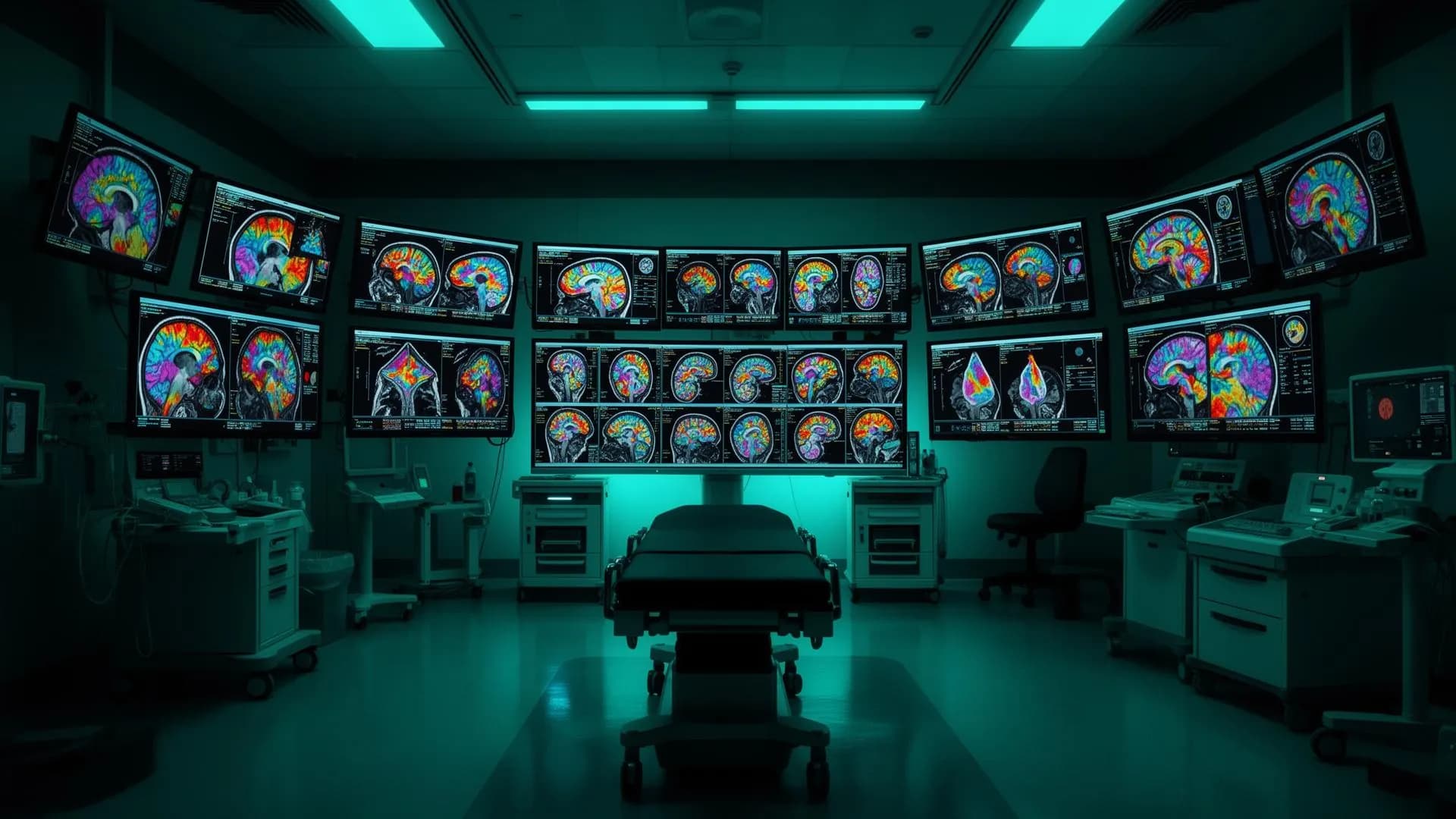
Advanced Tumor
Intelligence & Precision Imaging
See what standard scans can't. Advanced neuro-imaging maps your tumor's metabolic activity, vascular structure, and genetic markers, so your care team starts with the clearest possible picture of the disease.
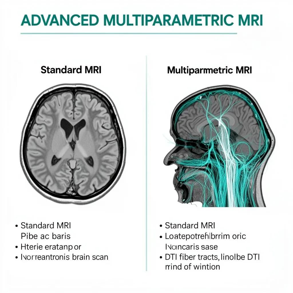
Why standard MRI isn't enough for GBM
A conventional MRI shows the tumor mass - but glioblastoma is infiltrative. Cancer cells extend far beyond what appears on a standard scan. Advanced multiparametric imaging reveals the full extent of disease, enabling more complete treatment planning and better outcomes.
Standard MRI
Shows mass only
DTI Mapping
Reveals fiber tracts
Perfusion MRI
Maps blood flow
PET Fusion
Metabolic activity
Advanced tumor intelligence & imaging
Precision treatment needs precise targets. Advanced neuro-imaging maps your tumor's metabolic activity, vascular structure, and genetic markers before treatment begins.
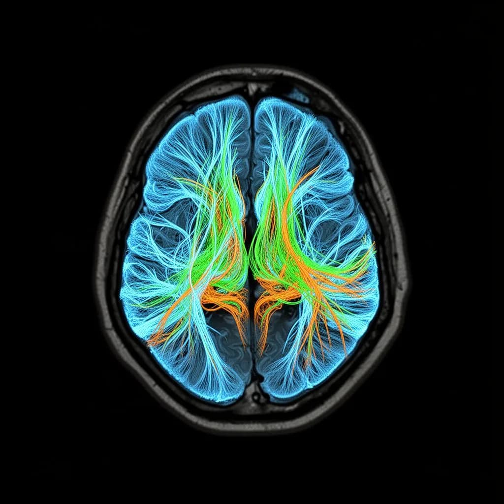
See the Invisible Pathways
Multiparametric MRI
Standard structural scans miss too much. DTI (Diffusion Tensor Imaging) and Perfusion MRI map the white matter tracts and blood flow around the tumor so the surgical and radiation plan spares healthy function.
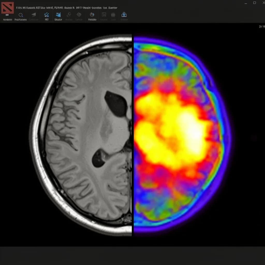
Molecular-Level Tumor Detection
DOTA PET/MR integration
By combining the high-resolution anatomy of an MRI with the metabolic mapping of a DOTA PET scan, we can identify specific cellular receptors. This allows us to pinpoint exact areas of active tumor growth that standard scans might miss.
The Virtual Biopsy
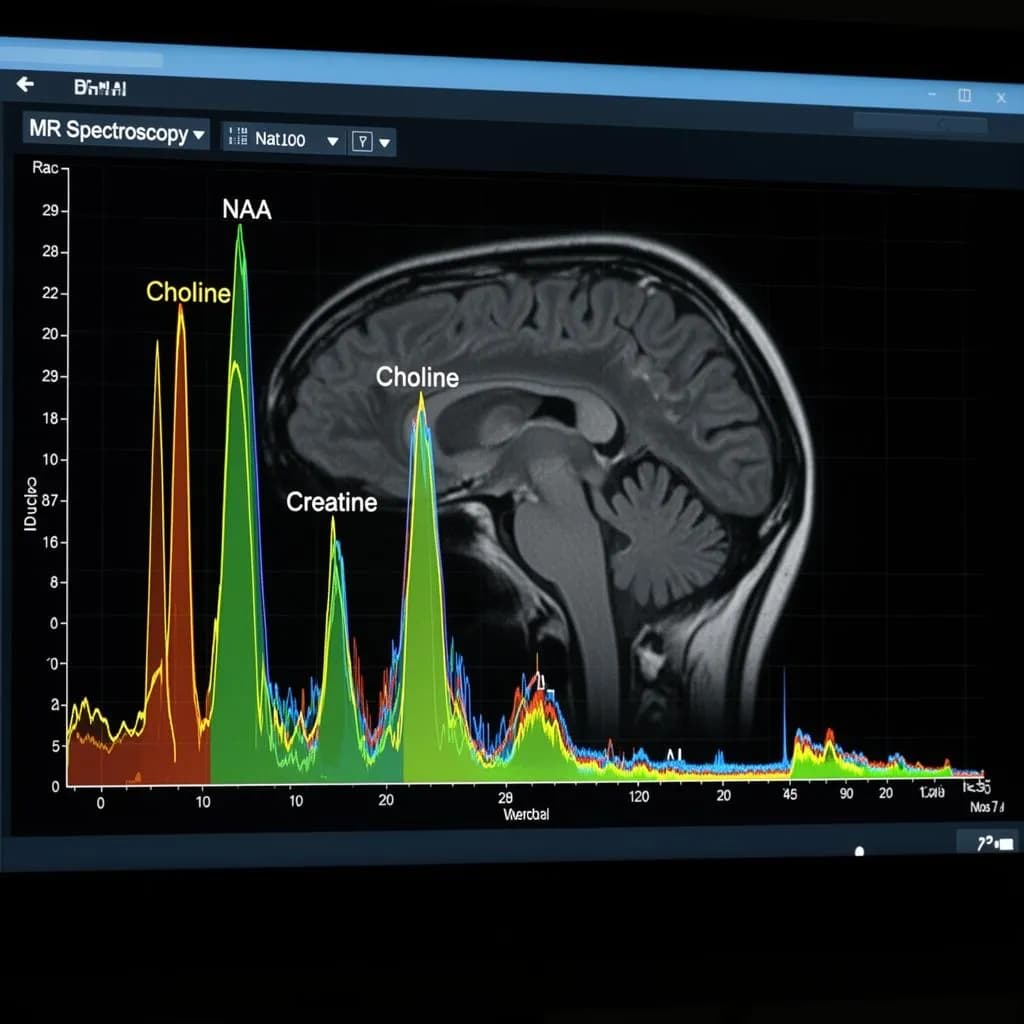
MR spectroscopy (MRS)
Tumors have a distinct chemical signature. MRS allows us to perform a "virtual biopsy" by analyzing the biochemical and metabolic profile of the tissue, helping us differentiate between tumor progression and treatment-related inflammation.
Precision Treatment Planning
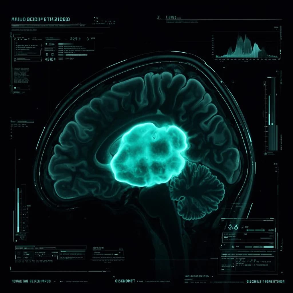
Amino acid PET imaging
Radiotracers map the amino acid metabolism of glioblastoma cells, informing targeted therapy choice and giving surgical and radiation plans a sharper target.
The GBM imaging challenge
Glioblastoma is notoriously difficult to image accurately. Its infiltrative nature means tumor cells extend well beyond what conventional MRI reveals, making advanced imaging not just helpful - but essential.
Infiltrative borders
GBM cells migrate along white matter tracts far beyond the visible tumor mass. Advanced DTI mapping reveals these hidden pathways.
Molecular heterogeneity
Different regions of the same tumor can have different genetic profiles. Multi-modal imaging helps identify these distinct zones for targeted therapy.
Treatment response
Distinguishing true tumor progression from pseudoprogression after treatment is critical. Spectroscopy and PET imaging provide the metabolic clarity needed.