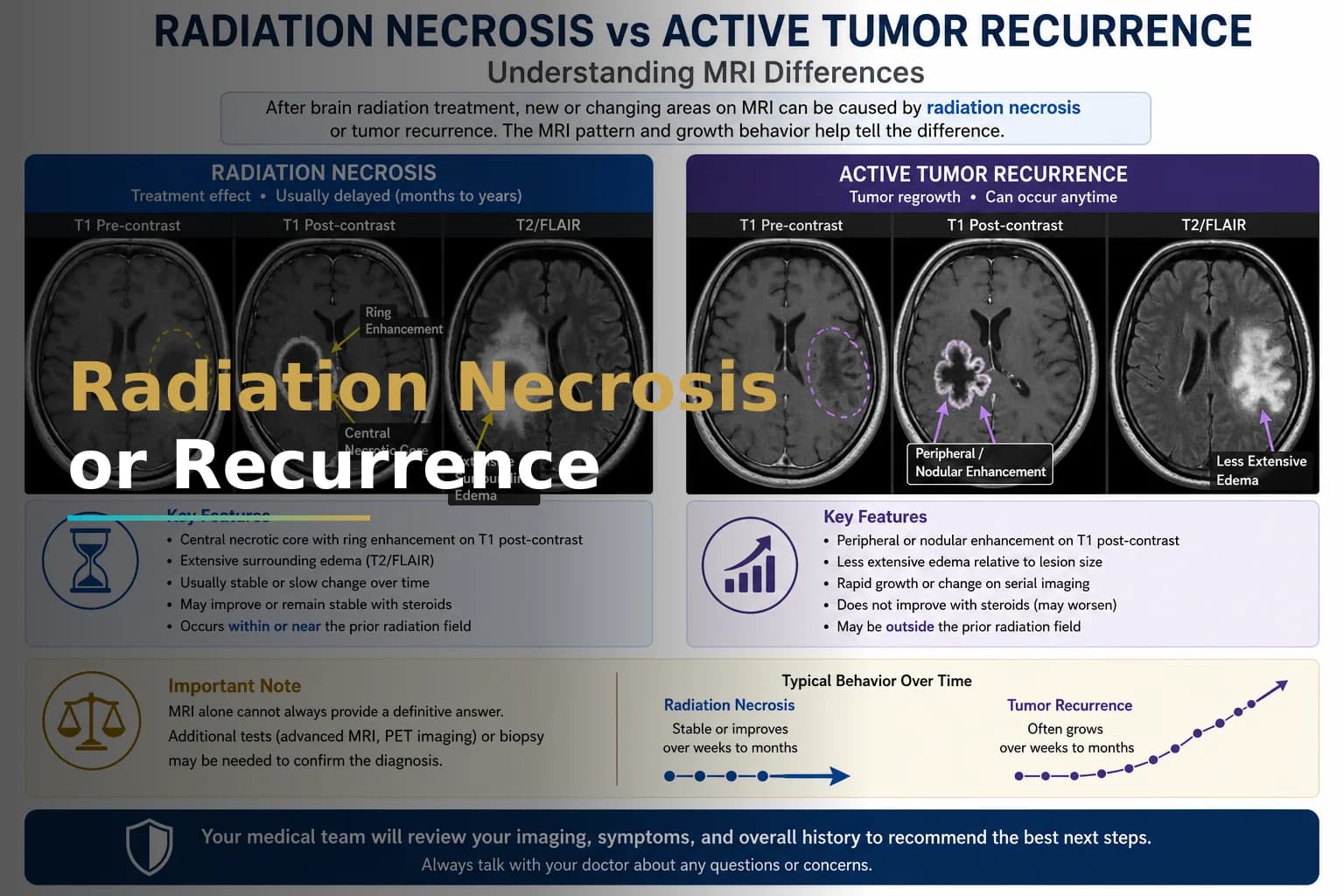
Radiation Necrosis vs Tumor Recurrence in Glioblastoma
A new or growing area on your MRI after glioblastoma treatment worries patients and caregivers. The question is almost always the same: is this the tumor coming back, or is it damage from the radiation? Getting the right answer matters because it changes what happens next.
What Is Radiation Necrosis After Glioblastoma Treatment?
Radiation necrosis is tissue damage that develops after radiation therapy to the brain. Healthy brain tissue in or near the treatment area dies in response to radiation. This dead tissue can swell, grow abnormal blood vessels, and appear on MRI scans just like a returning tumor does.
Radiation necrosis usually appears 3 to 12 months after radiation therapy ends, though it can happen as early as a few weeks or as late as several years later. According to PubMed Central, it happens in 3% to 24% of patients who get brain radiation. The amount depends on how much radiation was used, how much brain was treated, and whether chemotherapy was given too. Different hospitals use different practices, so the numbers vary.
Why This Difference Matters
If the abnormality is radiation necrosis but your doctors treat it as recurrence, you might get unnecessary chemotherapy, join a clinical trial based on wrong information, or get more radiation that damages your brain further. If it really is tumor coming back but your doctors treat it as necrosis, waiting for the right treatment costs time when the disease is moving fast.
This is not just a small detail. It is the difference between controlling inflammation and giving you second-line cancer treatment.
Symptoms: Why They Overlap
Radiation necrosis and tumor recurrence cause similar symptoms. Both can cause new or worse headaches, seizures, weakness or numbness, slower thinking, and tiredness. About half of patients with radiation necrosis notice these symptoms. The other half see changes on MRI but feel fine. You cannot tell them apart by symptoms alone.
If your team mentioned pseudoprogression - early swelling that appears during chemotherapy and radiation and can look like early recurrence - see the article on pseudoprogression in glioblastoma for details on that difference.
How Do Radiation Necrosis and Tumor Recurrence Differ on Imaging?
MRI scans can show both radiation necrosis and tumor recurrence, but they often cannot tell them apart by themselves. The table below shows the imaging patterns that doctors use to separate them.
| Feature | Radiation Necrosis | Tumor Recurrence |
|---|---|---|
| Typical timing after radiation | 3-12 months; delayed cases possible | Any time; most common after 6 months |
| Conventional MRI enhancement | Irregular, soap-bubble pattern; central low signal on T1 | Solid or nodular enhancement; rim-enhancing pattern common |
| Perfusion MRI (rCBV) | Low relative cerebral blood volume - minimal new vessel formation | High relative cerebral blood volume - actively growing vessels |
| MR spectroscopy (MRS) | Low choline; elevated lipid and lactate peaks; reduced NAA | Elevated choline-to-NAA ratio; high choline-to-creatine |
| Amino acid PET (FET or FDOPA) | Low or absent tracer uptake in affected area | Elevated tracer uptake matching active tumor metabolism |
| Response to corticosteroids | Lesion often shrinks or stabilizes on follow-up MRI | Minimal or no lasting response; lesion continues to grow |
Sources: Diagnosis and Treatment of Pseudoprogression, Radiation Necrosis and Brain Tumor Recurrence; Imaging Challenges for the Evaluation of Treated Gliomas
One scan alone won't give you the answer. The best approach uses DSC perfusion MRI with MR spectroscopy at least. Many major cancer centers also use amino acid PET, which gives a clearer picture than standard PET because normal brain tissue uses a lot of glucose that can hide the tumor signal. If your radiology report does not mention perfusion data or metabolite ratios, your workup might be incomplete.
Advanced Imaging Methods in More Detail
DSC Perfusion MRI
Dynamic susceptibility contrast (DSC) perfusion MRI measures relative cerebral blood volume (rCBV) inside a lesion. Tumors create new, leaky blood vessels to grow. Radiation necrosis does not. An rCBV ratio above about 2.0 compared to normal white matter suggests recurrence. A ratio below 1.5 suggests necrosis. These numbers change depending on the scan method and scanner type, which is why the same scan read at two different hospitals can give different answers. This is why getting a second opinion on imaging matters in unclear cases.
MR Spectroscopy
MR spectroscopy measures chemicals in brain tissue instead of showing its shape. Choline is a sign of active cell changes and is high in growing tumors. NAA shows healthy nerve cells and is low in both necrosis and tumors. A spectrum with high lipid and lactate peaks and low choline points to tissue death, not active tumor. Combined with perfusion data, spectroscopy greatly reduces the uncertainty, according to the Journal of Neuro-Oncology.
Amino Acid PET
Amino acid tracers like FET and FDOPA are taken up by actively dividing tumor cells but not by swollen or dead tissue. Several major European cancer centers now use amino acid PET as a standard step in the workup when standard MRI is unclear. Access varies: it is available in Germany, France, and parts of the US, but not as much in many other countries. If you cannot get this scan locally, getting it at a major cancer center before you decide on treatment makes sense.
When Imaging Still Cannot Tell the Difference
Even with advanced imaging, some cases stay unclear. When that happens, taking a tissue sample is the way to know for sure. Two procedures do this:
- Stereotactic biopsy - a needle biopsy guided by MRI, done through a small opening in the skull under anesthesia. It tells you exactly what the tissue is and is especially useful when the lesion is in a place where open surgery would be too risky.
- Laser interstitial thermal therapy (LITT) - a minimally invasive procedure where a laser fiber is placed inside the lesion under MRI and destroys the tissue with heat. For radiation necrosis, LITT works two ways - it shows you what the tissue is and removes the dead material at the same time. According to PubMed Central, surgery helped selected patients improve while also giving a clear answer about what was there.
If you are trying to decide whether to try salvage treatment, get a second opinion, or check clinical trial options before having an invasive procedure, see the guide on salvage treatment options for recurrent glioblastoma.
Treatment Options When Radiation Necrosis Is Confirmed
Treatment for radiation necrosis aims to reduce swelling and pressure on your brain without causing more damage. The treatment starts with the gentlest options and moves to stronger ones based on how you respond.
Corticosteroids
Dexamethasone is almost always the first treatment for radiation necrosis with symptoms. It can reduce brain swelling and improve symptoms in days. The problem is that long-term use has serious side effects - weight gain, high blood sugar, weak bones, and mood changes. Many patients keep needing steroids without the lesion going away.
Bevacizumab
Bevacizumab is an anti-VEGF (vascular endothelial growth factor) antibody. Radiation necrosis makes too much VEGF, which causes leaky blood vessels and swelling on MRI. Blocking VEGF can shrink the lesion and reduce swelling. According to PMC, bevacizumab improved both scans and symptoms in patients with radiation necrosis. A study in Neuro-Oncology Practice shows it works best in patients whose symptoms did not get better with steroids. Not all hospitals use it as a first treatment, and some areas have limited access.
Laser Interstitial Thermal Therapy
For lesions that do not respond to medicine, LITT is a less invasive surgical option. It needs MRI guidance and a neurosurgery team trained in it. LITT uses only a small hole in the skull instead of opening up the whole skull, so recovery is much faster than regular surgery. The procedure is available in a growing number of major centers in the US, UK, Germany, and some hospitals in India. If you cannot get LITT where you live, traveling to a specialist center for this treatment makes sense.
Hyperbaric Oxygen Therapy
Hyperbaric oxygen therapy (HBOT) gives high-pressure oxygen to tissues that do not get enough blood. There is limited evidence for HBOT in brain radiation necrosis - mostly case reports and small studies. According to StatPearls, most protocols use 20 to 40 sessions, which is a big commitment and is hard to find outside major cities. It is usually tried only when other options have not worked well enough.
Surgical Resection
Open surgery to remove dead tissue is used for patients with large mass effect, unclear diagnosis after all imaging, or disease that does not respond to other treatments. It gives the clearest answer about what is there and immediately relieves pressure. For patients who cannot have surgery because of where the lesion is or other health problems, the treatment options shrink a lot. This is why getting the imaging right early matters so much before choices get limited.
Accessing Expert Evaluation at International Centers
Getting the right interpretation of your scans often takes expertise that may not be at the hospital where you had treatment. This is especially true if your healthcare system does not do advanced perfusion MRI, does not have amino acid PET, or does not see many brain tumor cases. In those places, your local scan may not be read with the specific methods needed to use DSC perfusion to tell radiation necrosis from recurrence.
Major cancer centers in India, Germany, the US, and the UK have brain tumor imaging programs that do DSC perfusion MRI and MR spectroscopy regularly. Some of these centers also do LITT and have set up ways for international patients to get clear diagnosis before starting treatment at home. Having your scans reviewed by one of these programs without traveling is the most practical first step.
If you are not sure how to send your scans and medical records to a specialist in another country, see the guide on preparing for a remote glioblastoma second opinion. It explains what a specialist needs and how to send it. If you want help with your scans and do not want to do this alone, you can get a remote second opinion from Art of Healing Cancer. A specialist team will review your scans and reports and tell you if you need more testing or an in-person visit.
When to Talk to Your Doctor
Talk to your oncologist about radiation necrosis if your post-treatment MRI shows new or growing contrast areas in the radiation treatment area, especially if it appears 3 to 18 months after radiation ended and you have noticed neurological changes. Ask if DSC perfusion MRI or MR spectroscopy was done alongside the standard MRI and if the radiologist compared it to old scans. If the report does not mention perfusion or chemical markers, ask for an advanced imaging sequence or a second opinion on the imaging.
If you would like your scans and reports reviewed by a specialist team, you can upload them through the Glioblastoma Center patient journey page to request a structured remote case review.
This article is for general information and is not a substitute for medical advice. Always consult your oncologist or care team about your specific situation.