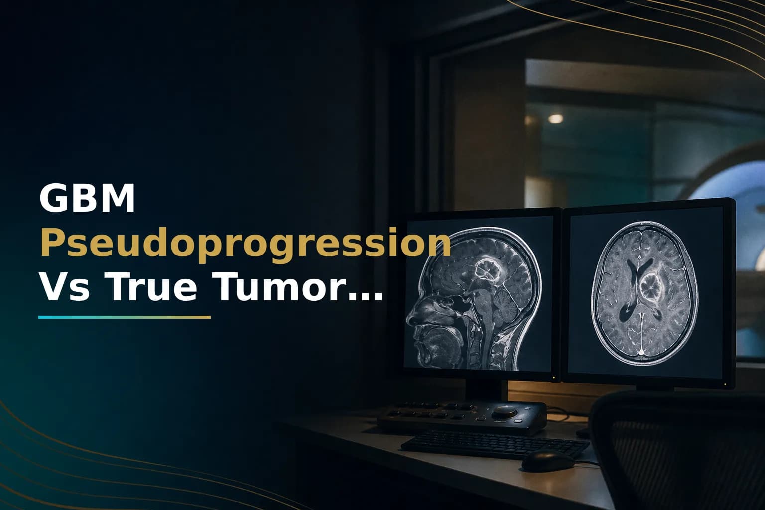
Pseudoprogression is a worrying but common finding in glioblastoma care. You finish six weeks of radiation and chemotherapy. A few weeks later, your MRI shows that the abnormal area has grown. Your team mentions possible tumor progression. But most often, the scan is showing something else: inflammation from treatment, not tumor growth. It's the brain's response to the damage radiation and chemotherapy caused.
What is pseudoprogression in glioblastoma?
Pseudoprogression is a treatment-related change on MRI that looks like tumor growth. It's actually inflammation from chemoradiation. It occurs in roughly 20 to 30 percent of glioblastoma patients after completing the Stupp protocol and usually goes away on its own without changing therapy. It does not mean your tumor has come back.
The clinical danger is real. If your team interprets pseudoprogression as true recurrence, they may stop or switch a treatment that is actually working. Understanding the difference matters. So does knowing when to ask for advanced imaging or a second opinion. Both are critical at this stage of care.
Why does pseudoprogression happen?
Radiation and temozolomide damage the blood-brain barrier, the network of cells that normally keeps large molecules out of brain tissue. After chemoradiation, that barrier becomes leaky. When radiologists inject contrast dye for an MRI, gadolinium seeps more freely into brain tissue than it normally would. The result is a bright patch on the scan that can look identical to an active, growing tumor.
At the same time, the immune system responds to radiation-killed cells by sending inflammatory signals to the treated area. Blood vessels dilate, fluid builds up, and local tissue swells. All of this increases contrast enhancement on imaging, even if there are no viable tumor cells actively dividing.
How common is pseudoprogression after chemoradiation?
A systematic review and meta-analysis of high-grade glioma studies found that between 20 and 36 percent of patients show increased contrast enhancement on the first post-radiation scan that later turns out to be pseudoprogression rather than true tumor growth. In some individual studies, the figure reaches as high as 50 percent, depending on the patient population and imaging criteria used.
The rate is high in the 12 weeks immediately after completing radiotherapy. Guidelines based on the Response Assessment in Neuro-Oncology (RANO) criteria recommend against labeling any new enhancement within that window as definitive progression. Unless there is histologic evidence of active tumor, or the patient shows clear clinical decline while off corticosteroids, the standard approach is to repeat imaging and wait before changing treatment.
Does your MGMT methylation status affect your risk?
Yes, significantly. MGMT promoter methylation tells clinicians whether temozolomide is likely to work against your specific tumor. It also predicts how likely you are to develop pseudoprogression. Research published in the Journal of Clinical Oncology found that pseudoprogression after concurrent chemoradiation occurred more often in patients with a methylated MGMT promoter than in those with an unmethylated one.
This makes biological sense. MGMT-methylated tumors respond better to temozolomide, meaning the drug does more damage to tumor cells and the surrounding blood-brain barrier. More treatment effect means more treatment-related inflammation and a higher chance that the first post-treatment scan will show changes that resemble true progression.
If you don't yet know your MGMT status, this is one important reason to find out before your first follow-up MRI. A molecular testing report completed in the weeks after surgery shapes not only your initial treatment plan but also how you and your care team should interpret every scan that follows.
When does pseudoprogression typically appear?
Most cases show up within the first 12 weeks after completing chemoradiation, often at the very first follow-up MRI. Neuro-oncologists have traditionally used this window as a guide: enhancement that appears and then stabilizes or shrinks within 12 weeks, without any change in treatment, is more likely to be pseudoprogression than true growth.
That timing is not a hard rule. A case series published in Frontiers in Oncology documented delayed pseudoprogression in glioblastoma patients receiving Tumor Treating Fields (TTFields/Optune), where radiographic worsening appeared six months or more after completing radiation. The proposed mechanism is that TTFields increase cell membrane permeability, allowing more gadolinium contrast to leak into brain tissue and create MRI changes that mimic progression well beyond the standard 12-week window. If TTFields are part of your maintenance therapy, your team should keep delayed pseudoprogression on the differential even at later scan time points.
For more details on how TTFields work and what to expect across months of maintenance therapy, see our guide to Tumor Treating Fields and Optune for glioblastoma.
How do doctors distinguish pseudoprogression from true recurrence?
Standard contrast-enhanced MRI cannot reliably make this distinction on its own. Both pseudoprogression and true tumor recurrence produce new or enlarged areas of enhancement, and both can worsen symptoms in some patients. Drawing on systematic reviews of metabolic and functional MRI in glioblastoma, most specialist centers now use a combination of advanced imaging techniques to build a more complete picture.
Perfusion MRI
Perfusion MRI measures blood flow and blood volume in brain tissue. Tumors are metabolically active and build new, abnormal blood vessels to sustain themselves, a process called angiogenesis. True tumor recurrence therefore tends to show high relative cerebral blood volume (rCBV). Pseudoprogression, driven by inflammation rather than active proliferation, typically shows lower rCBV values. Dynamic susceptibility contrast (DSC) perfusion imaging is the most widely used technique for this purpose and improves diagnostic accuracy beyond standard MRI alone.
MR spectroscopy
Magnetic resonance spectroscopy (MRS) maps the chemical composition of brain tissue without a biopsy. Active tumor cells produce elevated choline, a marker of rapid cell membrane turnover, alongside reduced N-acetylaspartate, a marker of healthy neurons. Inflamed tissue shows a different metabolic profile. A 2022 study found that MRS outperformed perfusion imaging in distinguishing pseudoprogression from true disease progression in glioblastoma patients, with the choline-to-N-acetylaspartate ratio demonstrating the highest diagnostic accuracy in the cohort studied.
Diffusion-weighted imaging
Diffusion-weighted imaging (DWI) measures how freely water molecules move through tissue. Densely packed, dividing tumor cells restrict water movement, producing lower apparent diffusion coefficient (ADC) values. Inflammatory tissue from pseudoprogression allows more free water movement and typically shows higher ADC values. DWI is widely available and adds meaningful data when interpreted alongside perfusion and spectroscopy results.
Amino acid PET
Positron emission tomography using amino acid tracers - particularly FET-PET, which labels a synthetic amino acid rather than glucose - has shown promise in separating tumor from treatment-related changes. Tumor cells absorb amino acids at higher rates than inflamed tissue. FET-PET is currently being evaluated in randomized clinical trials specifically aimed at pseudoprogression management in glioblastoma patients. Standard FDG-PET is less useful in brain tumors because healthy brain tissue has high baseline glucose uptake, making it difficult to detect tumor signal against that background.
What about a tissue biopsy?
When imaging remains unclear and the clinical stakes are high, a repeat biopsy or surgical re-resection can provide more definitive information. A pathologist examining the tissue directly can assess whether the sample contains actively dividing tumor cells, treatment-related necrosis, or a mix of both. The tradeoff is the risk of any brain surgery and the fact that pseudoprogression and tumor recurrence can coexist in the same specimen, which sometimes makes even pathology results complex to interpret.
Surgery is generally considered when neurological symptoms are worsening significantly, when imaging remains unclear after repeat scanning, or when re-resection might itself be clinically beneficial. This decision benefits from multidisciplinary review at a center that manages a high volume of glioblastoma cases.
What to do when your scan result is ambiguous
First: an MRI showing new or enlarged enhancement after chemoradiation does not automatically mean treatment has failed. Ask your oncologist directly whether the finding meets the criteria for possible pseudoprogression - specifically, whether it appeared within 12 weeks of completing radiation, without clear clinical decline, and without biopsy-proven tumor.
Second, ask whether advanced imaging is available at your center. Perfusion MRI and MR spectroscopy are not universally available, but most specialist neuro-oncology centers can perform them. If your scan is being read at a center without dedicated neuroradiology expertise, requesting a second read from a specialist neuroradiologist is entirely reasonable.
Third, if the interpretation remains unclear and your team is considering a treatment change based on a single unclear scan, seek a second opinion before acting. A neuro-oncologist reviewing the same imaging alongside your clinical history and MGMT methylation status may reach a very different conclusion. Stopping an effective treatment during pseudoprogression could mean abandoning a therapy that is still working.
Facing an unclear scan is frightening, and you should not have to work through it alone. If you want experienced eyes on your MRI and pathology reports before making any decision, you can arrange a remote second opinion through Art of Healing Cancer, whose team specializes in reviewing GBM imaging cases for patients and caregivers in exactly this situation.
For a full breakdown of what a glioblastoma second opinion covers - including which imaging studies and molecular reports should be included - see the glioblastoma second opinion checklist. To have your full case reviewed before any treatment decision, you can upload your MRI and reports through the Glioblastoma Center patient journey form to request a remote case review.
When to talk to your doctor
Contact your oncology team promptly if new or worsening neurological symptoms appear between scheduled scans: increased headaches, new weakness, speech changes, or seizures. These need evaluation regardless of whether pseudoprogression is under consideration. If your next MRI is more than six to eight weeks away and your condition is changing, request an earlier scan rather than waiting.
If your team is recommending a treatment change based on a single unclear scan taken within 12 weeks of completing chemoradiation, it is reasonable to ask: could this be pseudoprogression, and should we repeat the imaging in four to six weeks before making any change?
This article is for general information and is not a substitute for medical advice. Always consult your oncologist or care team about your specific situation.