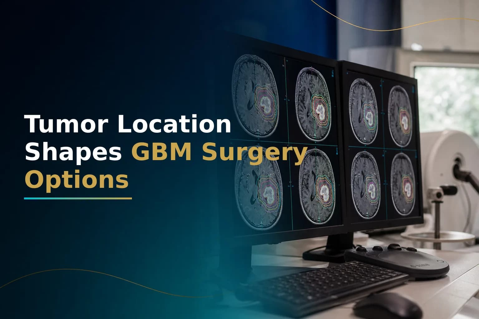
How Glioblastoma Tumor Location Shapes Surgery Options
Where a glioblastoma grows in the brain matters as much as the diagnosis itself. Location determines whether your surgeon can safely remove most of the tumor, what techniques they'll use, and how the radiation team plans your treatment. Two patients with a grade 4 glioblastoma can face very different surgery options depending on whether the tumor is in the front of the brain or deep near structures that control movement, speech, or vision. This guide helps you ask better questions and decide if a second opinion is worth getting.
What does tumor location mean for treatment?
Tumor location shapes two key decisions. First is how much tumor can be safely removed and what technique the surgeon will use. Second is how tightly radiation can be aimed without damaging nearby structures. The frontal lobe gives surgeons the most options. Deep structures like the thalamus and corpus callosum often limit surgery to a needle biopsy. Most other locations fall somewhere between these extremes.
How does tumor location change what surgery is possible?
| Consideration | Frontal / Non-Eloquent | Eloquent or Deep-Brain | Corpus Callosum (Butterfly) |
|---|---|---|---|
| Typical surgery goal | Maximal safe resection; gross total removal often achievable | Partial resection or biopsy; awake craniotomy used to protect function | Stereotactic biopsy in most cases; full resection rarely feasible |
| Key surgical technique | 5-ALA fluorescence-guided craniotomy under general anaesthesia | Awake craniotomy with cortical mapping; or stereotactic needle biopsy | Stereotactic needle biopsy |
| Radiation margin complexity | Lower - standard IMRT margins usually feasible | Higher - proximity to brainstem, optic tracts, or language areas constrains dose | Very high - bilateral involvement makes standard margins difficult to plan |
| Proton therapy relevance | Moderate - may reduce dose to healthy brain tissue | Stronger case, particularly for thalamic or brainstem-adjacent tumors | Assessed case by case; bilateral spread complicates beam planning |
| Primary functional risk | Relatively lower in anterior frontal regions | Motor, language, sensory, or visual deficits possible | Cognitive and motor deficits; bilateral damage risk |
Better locations - meaning farther from areas that control critical functions - offer more surgery options and simpler radiation planning. Harder locations do not mean no options. They mean your team needs more advanced techniques and a more detailed conversation about risk and benefit before deciding on a plan.
Frontal lobe tumors: the most surgically flexible territory
The frontal lobe has two broad zones. The posterior frontal region - particularly the primary motor cortex that drives movement, and Broca's area (which controls speech production in most right-handed people) - is considered eloquent. Damage here causes lasting deficits. The anterior frontal region has more functional overlap and redundancy. Losing tissue here more often causes subtle changes in personality or planning ability rather than paralysis or permanent speech loss.
When a glioblastoma sits in the non-eloquent anterior frontal area, neurosurgeons can usually pursue aggressive removal. Fluorescence-guided surgery using a compound called 5-ALA (5-aminolevulinic acid) is a key tool. The patient takes an oral dose a few hours before surgery. The compound builds up in tumor cells and glows pink under a blue-violet surgical light, helping the surgeon see tumor borders that would otherwise look like normal brain tissue. Multiple trials show this technique increases the rate of complete removal of the contrast-enhancing tumor.
A systematic review and meta-analysis confirmed that increasing extent of resection is associated with longer overall survival in IDH-wildtype glioblastoma - the most common form of this tumor. For a detailed breakdown of that survival evidence, see How Glioblastoma Extent of Resection Affects Survival.
For tumors that extend into or near the eloquent posterior frontal region, the surgeon will discuss either a more limited resection or an awake craniotomy - described below.
Temporal lobe tumors: language is the central concern
A glioblastoma in the temporal lobe raises a specific concern. In the vast majority of right-handed people, and many left-handed people, the left temporal lobe contains Wernicke's area - the region that processes language comprehension. Damaging Wernicke's area causes a form of aphasia where a person can produce fluent-sounding speech but it carries little meaning, and they struggle to understand what others say.
Surgeons operating on a left temporal lobe tumor almost always consider awake craniotomy with language mapping to identify and protect that region before removing any tissue near it. Right temporal lobe tumors carry lower language risk and allow more surgical flexibility. In some specialized centers, surgeons discuss a more extensive operation called anterior temporal lobectomy - removing a larger block of temporal tissue - where emerging evidence suggests this may improve seizure control and possibly extend survival in carefully selected patients.
Parietal and occipital lobe tumors: sensation and vision at stake
The parietal lobe processes sensory information and spatial awareness. A glioblastoma near the sensorimotor strip can affect sensation or fine coordination on one side of the body. These tumors often call for awake craniotomy with both motor and sensory mapping, or careful preoperative planning using functional MRI (fMRI) and diffusion tensor imaging (DTI) - techniques that map brain function and major fiber tracts on a scan before surgery.
Occipital lobe tumors are less common. They sit near the visual cortex, and resection here risks affecting the visual field. The surgical team will map visual function carefully, and the radiation oncologist must plan around the optic nerves and optic tracts to protect vision. Some patients with occipital tumors notice changes in their visual field at the time of diagnosis.
What an awake craniotomy actually involves
Awake craniotomy is often misunderstood. Pain is not the concern it may sound like - the scalp and skull are numbed with local anesthetic, and the brain itself has no pain receptors. The patient stays alert and cooperative during the part of the procedure when the surgeon maps the brain surface. They might count aloud, name objects, or perform a simple movement task while the surgeon delivers brief, low-level electrical pulses to small patches of exposed cortex. If a pulse causes a word to get stuck or a hand movement to stall, that area is marked and avoided.
A systematic review and meta-analysis of awake craniotomy for glioblastoma found that doctors achieved gross total resection of 95% or more in 64% of procedures, and resection of 85% or more in 77% - meaningful rates for tumors in eloquent territory that would otherwise be approached far more conservatively. Neurological worsening appeared in about 21% of patients whose mapping identified eloquent cortex, compared to around 9% in those whose mapping was negative. Many of these changes were temporary and improved as post-surgical swelling went down over days to weeks.
Not every patient is a candidate. Pre-existing cognitive difficulty, severe anxiety, or a tumor position that makes the required positioning intolerable can rule it out. This is exactly where a second neurosurgical opinion at a center that performs awake craniotomy regularly can clarify what is technically feasible for the specific anatomy involved.
Deep-brain and midline tumors: when surgery becomes limited
Tumors in the thalamus, basal ganglia, or brainstem occupy territory densely packed with pathways controlling movement, sensation, consciousness, and basic body functions. Attempting standard surgery here risks severe, irreversible damage. Surgery in these locations is generally limited to a stereotactic needle biopsy - a minimally invasive procedure where a thin needle guided by MRI coordinates reaches the tumor through a small hole in the skull and extracts tissue for diagnosis and molecular profiling. Most patients go home within a day or two.
That biopsy still provides critical information: MGMT methylation status, IDH mutation testing, and other molecular markers that guide chemotherapy choices and clinical trial eligibility. A study of biopsy-only glioblastoma patients found that a subset with favorable molecular profiles showed extended survival - confirming that biopsy information is a real treatment-planning tool, not just information when surgery isn't possible.
Because surgery is limited for deep tumors, radiation becomes the primary treatment alongside chemotherapy. Research comparing IMRT to older 3D conformal techniques found that IMRT reduced the percentage of brainstem volume receiving doses above 45 Gy by 31% - meaningful protection when the tumor sits close to that structure. For tumors adjacent to the brainstem or optic apparatus, the radiation team may also evaluate proton therapy, which deposits most of its energy at the tumor target and drops off sharply beyond it, reducing incidental dose to adjacent critical structures. For a detailed breakdown of when proton therapy makes clinical sense, see Proton Therapy for Glioblastoma: When It Is Indicated and How It Differs from Standard Radiation.
For tumors that have come back in a deep or previously irradiated region, laser interstitial thermal therapy (LITT) - also called laser ablation - has emerged as a minimally invasive option that does not require open craniotomy. The comparison of laser ablation versus re-irradiation for recurrent glioblastoma covers the evidence and patient eligibility criteria in detail.
Corpus callosum butterfly glioblastoma
When a glioblastoma crosses the corpus callosum - the thick band of nerve fibers connecting the two brain hemispheres - and infiltrates both sides simultaneously, imaging shows what neuro-oncologists call a butterfly pattern. The tumor mirrors itself on each side of the midline. Bilateral involvement of this kind typically makes gross total resection unsafe or impossible: removing enough tissue from both hemispheres would cause severe, lasting cognitive and motor damage.
In most butterfly GBM cases, the team proceeds with a stereotactic biopsy to confirm the diagnosis and obtain molecular markers, then moves to radiation and temozolomide chemotherapy on the standard Stupp protocol. A surgical state-of-the-art review notes that patient selection and clear goal-setting are as central to outcomes in these complex cases as technical expertise. Clinical trial enrollment becomes especially important in this group, where surgery cannot reduce the tumor as much as in single-hemisphere cases.
What to ask your team based on your tumor's location
If your MRI report or surgical consult mentions eloquent cortex involvement, midline extension, thalamic involvement, or bilateral spread, these are the specific questions worth asking before any consent form is signed:
- What extent of resection are you aiming for, and why?
- Is awake craniotomy an option here, and how often does your center perform it for glioblastoma?
- What are the realistic risks to speech, movement, or vision if a small margin of error is crossed?
- Has 5-ALA fluorescence guidance or intraoperative MRI been evaluated for my case?
- Does my radiation plan include specific measures to protect the brainstem, optic tracts, or language cortex?
- Is proton therapy worth evaluating given where my tumor sits?
If the answers feel incomplete or the center does not offer advanced mapping techniques, getting input from a higher-volume neuro-oncology center is a reasonable next step - and it does not have to mean traveling abroad on a tight timeline. You can arrange a remote second opinion through Art of Healing Cancer to have your MRI and surgical plan reviewed by an experienced team before committing to any approach.
If you want to map out how your specific tumor location affects your full range of options, you can upload your MRI and pathology reports on the Glioblastoma Center patient-journey page to request a remote case review from the team.
When to talk to your doctor
Seek a direct conversation with your neurosurgeon if your report names eloquent cortex, deep-brain structures, or bilateral extension and a clear surgical plan has not been explained. Ask specifically whether fluorescence-guided surgery, awake craniotomy, intraoperative MRI, or proton radiotherapy has been evaluated for your situation. If your center does not offer those techniques, ask for a referral to a specialist center that does. Each tool exists precisely for situations where standard approaches reach their limits.
This article is for general information and is not a substitute for medical advice. Always consult your oncologist or care team about your specific situation.