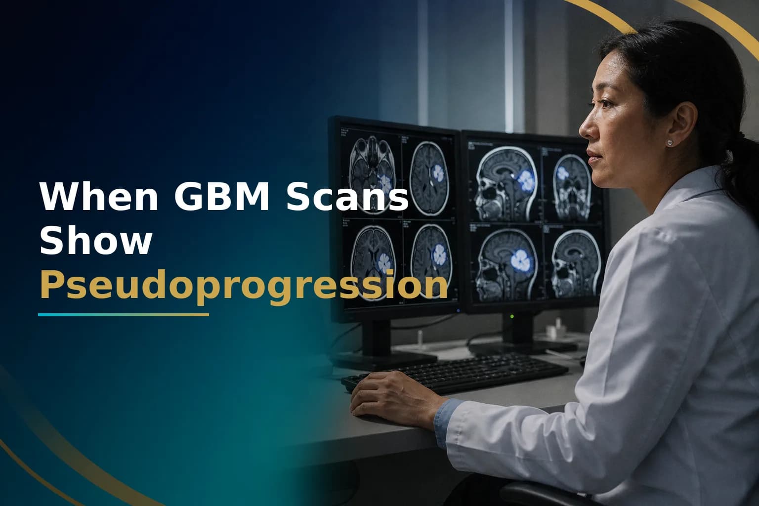
If the MRI taken a few weeks after glioblastoma chemoradiation shows new or bigger areas of enhancement, the immediate instinct is fear. Those changes can look exactly like tumor growth. But in a significant number of patients, they are not growth at all. They are a treatment effect called pseudoprogression, and distinguishing one from the other drives some of the most important clinical decisions in GBM care.
What Is Pseudoprogression in Glioblastoma?
Pseudoprogression is a worsening of brain MRI findings - new or enlarged areas of contrast enhancement - that appears after chemoradiation but does not reflect actual tumor growth. It typically occurs within the first 12 weeks after completing radiotherapy and temozolomide. The changes usually stabilize or improve without any change to the treatment plan.
According to a systematic review on metabolic and functional MRI in glioblastoma, pseudoprogression affects approximately 9% to 30% of patients treated with standard chemoradiation. That range varies because different centers use different definitions and imaging protocols. But the point is clear: a worrying scan after treatment does not automatically mean treatment failed.
Why Does Pseudoprogression Happen?
During chemoradiation, radiation temporarily disrupts the blood-brain barrier - the lining that controls what passes between the bloodstream and brain tissue. When that barrier breaks down, fluid leaks in and surrounding tissue becomes inflamed. Temozolomide can intensify this process by directly damaging local cells. The result is swelling and increased permeability that shows up as new enhancement on MRI, even though no new tumor cells are causing it.
This differs from true progression, where actively dividing tumor cells invade surrounding brain tissue and generate new blood vessels. On standard gadolinium-enhanced MRI, the two processes look nearly identical. That is the core diagnostic challenge.
Who Is More Likely to Experience Pseudoprogression?
The strongest known predictor is MGMT promoter methylation status. MGMT is a DNA-repair gene. When its promoter is methylated (switched off), tumor cells cannot repair the DNA damage caused by temozolomide as effectively. That greater sensitivity to chemotherapy appears to trigger a stronger local inflammatory response - and a higher rate of pseudoprogression on imaging.
In an early landmark study referenced in PubMed, pseudoprogression occurred in approximately 91% of patients with a methylated MGMT promoter, compared to 41% in those with unmethylated status, after concurrent radiochemotherapy. The sample in that original study was small, but later research confirmed the link between MGMT methylation and higher pseudoprogression rates. MGMT-methylated patients tend to have better overall outcomes from standard treatment - which makes correct identification of pseudoprogression especially important in this group. Stopping or changing treatment prematurely based on an ambiguous scan could mean abandoning a regimen that is actually working.
Understanding your molecular markers matters. The article What Your Glioblastoma Pathology Report Really Means explains how to read MGMT status and other key molecular findings from your neuropathology report in plain terms.
Why Standard MRI Cannot Always Separate the Two
Gadolinium-enhanced MRI shows where the blood-brain barrier is disrupted. That disruption occurs in both true progression (where growing tumor breaks down surrounding vasculature) and pseudoprogression (where treatment-related inflammation disrupts the same barrier). Standard MRI shows the effect but not its cause.
This is why a single scan showing an enlarging bright area after treatment cannot diagnose recurrence on its own. The image shows a biological signal - blood-brain barrier breakdown - but not what caused it. You need additional diagnostic tools, and sometimes repeated imaging, to tell the two apart.
How Do Doctors Distinguish Pseudoprogression from True Progression?
Several advanced imaging approaches can provide more information than standard MRI alone:
- Perfusion MRI (DSC or ASL): Measures relative cerebral blood volume (rCBV) inside the lesion. Tumors generate new blood vessels, producing high rCBV. Pseudoprogression typically shows lower rCBV. According to a review on pseudoprogression versus true progression in glioblastoma, perfusion MRI is the most widely used technique in routine clinical practice for this purpose.
- MR Spectroscopy (MRS): Measures tissue chemical composition. Active tumor tends to show elevated choline (a marker of cell membrane turnover) and reduced N-acetylaspartate, or NAA, a marker of healthy neurons. Research published in Neuro-Oncology Advances found that MRS may outperform perfusion MRI in diagnostic accuracy, though it is technically difficult and not available at every hospital.
- Amino acid PET (FET-PET): Uses a radioactive tracer absorbed by metabolically active tumor cells. It can help identify active tumor within areas where standard MRI findings are unclear. Availability varies widely by institution and country.
- Serial MRI over time: Often the most practical approach. True progression usually grows on follow-up scans over 4-8 weeks. Pseudoprogression usually stops growing or shrinks while you keep the same treatment.
- Biopsy: Shows for certain whether the area contains treatment-related necrosis and inflammation or actively dividing tumor cells. Biopsy has some risk and isn't always needed, but it's an option when you need a clear answer.
What the RANO 2.0 Criteria Say
The Response Assessment in Neuro-Oncology (RANO) criteria provide the international standard for defining response and progression in glioma. RANO 2.0, referenced in ASCO Publications, addresses pseudoprogression directly.
In newly diagnosed patients, the post-radiotherapy MRI - not the post-surgical MRI - serves as the reference baseline for comparison. Within the first 12 weeks after completing radiotherapy, when pseudoprogression risk is highest, RANO 2.0 states that true progression exists only if new enhancement is clearly outside the radiation field, or if a biopsy shows active tumor growth. In all other cases, you continue treatment and get a repeat MRI to confirm the finding before concluding the tumor has progressed. This framework prevents doctors from changing treatment too early based on unclear scans.
What Happens to Treatment During This Period?
When pseudoprogression cannot be confirmed or ruled out, the standard approach is to continue your current treatment plan and repeat the MRI at the next scheduled appointment - typically 6-8 weeks later. If the area continues to expand significantly on that repeat scan, or if you develop clear new neurological problems, this suggests true progression, and your doctor may change your plan.
Steroid dosing (typically dexamethasone) may be adjusted to manage swelling and symptoms during this time. Unnecessary increases in steroids carry their own side effects, so the goal is careful management while waiting for more scans to show what is happening.
Waiting without answers is genuinely hard for patients and the people doing research on their behalf. If your hospital does not have perfusion or spectroscopy MRI, or if two consecutive scans did not show what is really happening, ask a specialist to review your case. You can have the pathology and MRI reviewed by the Art of Healing Cancer team, who can assess whether more detailed imaging or a remote specialist consultation would give you clear answers about what to do next.
How This Connects to Evaluating First-Line Treatment Response
Pseudoprogression is one part of a broader question that arises after completing the first phase of chemoradiation: how should any MRI finding be interpreted at this stage, and what counts as a genuine response versus a failure? The article Evaluating Glioblastoma Response After First Chemotherapy addresses that full picture, showing how imaging, clinical signs, and genetics together show what response means and what unclear findings typically mean for next steps.
A closely related but distinct phenomenon is radiation necrosis - where radiation-damaged tissue causes persistent enhancement that can appear months to years after treatment, well beyond the early post-treatment window typical of pseudoprogression. Unlike pseudoprogression, radiation necrosis often lasts longer and may need treatment instead of waiting. The article Radiation Necrosis vs Tumor Recurrence in Glioblastoma explains how these two conditions are separated on advanced imaging and in the clinic.
Questions to Ask Your Neuro-Oncologist
- Based on my MGMT status, how likely is pseudoprogression in my specific case?
- Does this center have access to perfusion MRI or MR spectroscopy, and would you recommend either for my scan?
- What specific features on this scan concern you most, and what would you expect to see on the next scan if this is pseudoprogression rather than true progression?
- If the next scan also shows worsening, at what point would biopsy make sense rather than continued observation?
- What symptoms should prompt me to call before my next scheduled appointment?
When to Talk to Your Doctor
Contact your care team promptly if you notice sudden new weakness on one side of the body, rapidly worsening headaches, new or increased seizure activity, significant confusion, or abrupt changes in speech or vision between scheduled appointments. These symptoms may reflect pseudoprogression, true progression, or other treatment-related effects - they need immediate medical attention, not just waiting for your next MRI. Do not rely on just one scan to make a decision, especially during the first 12 weeks after completing chemoradiation.
If you're not sure whether the right imaging tools are being used to evaluate your post-treatment scans, you can send your MRI reports and treatment summary to the Glioblastoma Center patient journey form for review.
This article is for general information and is not a substitute for medical advice. Always consult your oncologist or care team about your specific situation.