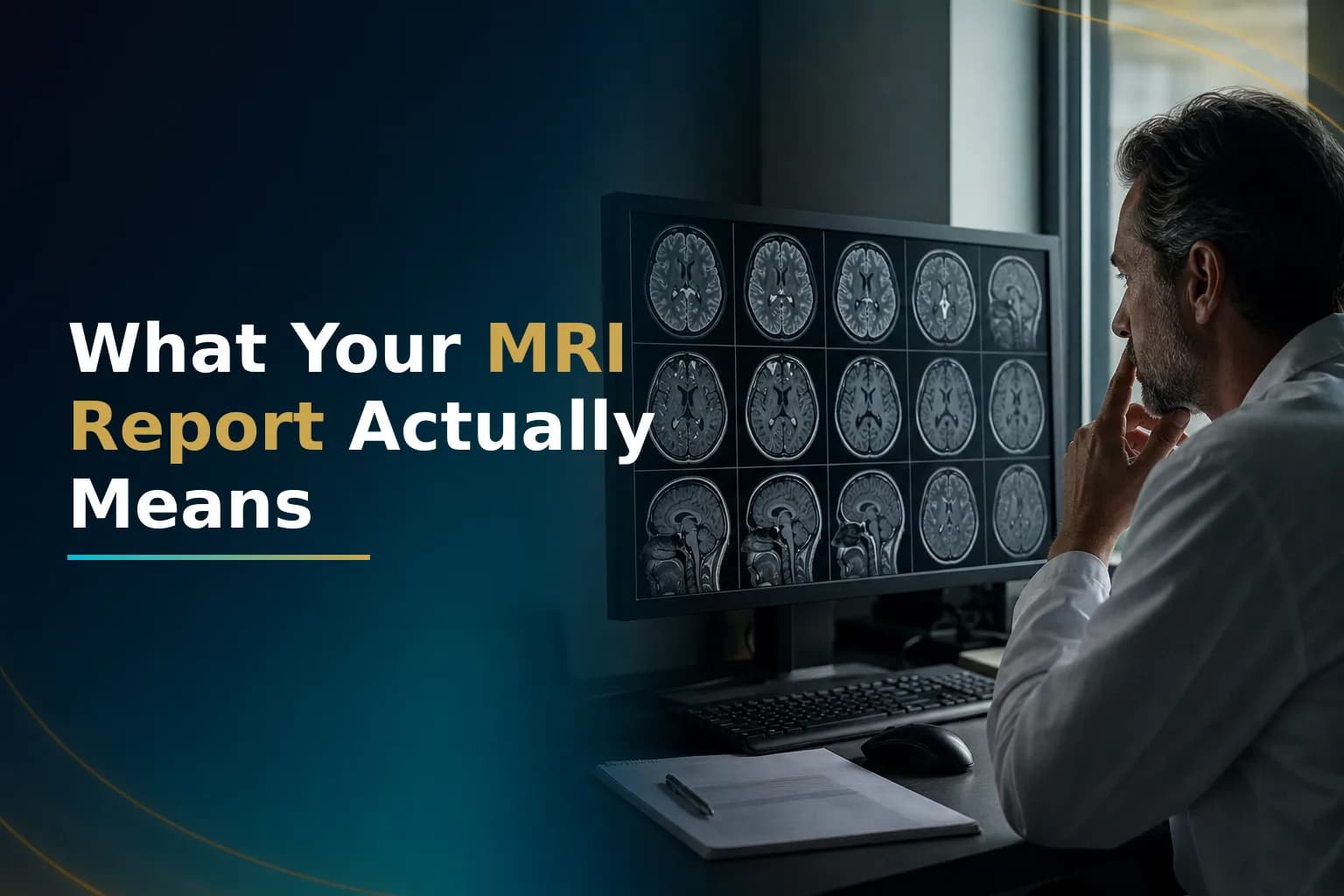
The MRI report arrives a few days after the scan. You scroll through medical shorthand and anatomy terms looking for one sentence that explains what is happening inside the tumor. Most patients and caregivers find radiology reports nearly impossible to read without a guide. That is not a failure of intelligence - it is a failure of translation.
This guide walks through the most common terms in a glioblastoma MRI report. You can follow the radiologist's reasoning and come prepared to your next oncology conversation.
What does a glioblastoma MRI report tell you?
A glioblastoma MRI report describes the tumor's size, location, and shape across multiple scan types called sequences. Each sequence highlights a different tissue property. Together they show how active the tumor is and how much pressure it places on the brain. Comparing scans over time also matters. One MRI alone is not the whole story - change over time matters as much as any single reading.
The standard MRI sequences and what each one shows
T1-weighted imaging
T1 is the baseline structural scan. Normal brain tissue appears in shades of gray. Fatty tissue looks bright. Fluid-filled spaces like the ventricles appear dark. The ventricles are chambers that hold cerebrospinal fluid. On T1 without contrast, glioblastoma typically appears slightly darker than surrounding brain tissue. This helps the radiologist map the tumor's boundaries.
T1 with gadolinium contrast (T1+Gd)
This is the most critical sequence for tracking GBM. Before the scan, a dye called gadolinium is injected into a vein. In a healthy brain, gadolinium stays inside blood vessels. In a tumor, the blood-brain barrier is disrupted. This is the tight lining that controls what enters brain tissue. Gadolinium leaks through and appears as a bright white signal on the scan.
That bright signal is called contrast enhancement. In GBM, it often forms an irregular bright ring around a darker center. The center is dead tissue inside the tumor. This pattern is described as a ring-enhancing lesion and is one of the hallmark imaging findings in glioblastoma, as documented in the NIH StatPearls reference on glioblastoma multiforme.
T2-weighted imaging
T2 makes water-rich tissue appear bright. Swelling and tumor infiltration both increase water in tissue, so they both appear bright on T2. This sequence shows how the tumor and surrounding inflammation are affecting the brain.
FLAIR (Fluid-Attenuated Inversion Recovery)
FLAIR is a modified T2 sequence that suppresses the signal from normal cerebrospinal fluid. This makes the ventricles appear dark. The suppression makes abnormal tissue stand out more clearly, especially near the ventricles. In GBM, FLAIR typically shows a larger area of abnormality than the contrast-enhancing region. That broader bright zone reflects a mix of swelling and tumor cells that have spread into surrounding brain tissue. These cells have not fully broken down the blood-brain barrier.
Serial changes in the T2/FLAIR area can signal tumor spread. New T2/FLAIR lesions away from the primary tumor cavity may indicate disease spread, as noted in research on progression patterns. These lesions need close attention on follow-up scans.
DWI (Diffusion-Weighted Imaging)
DWI measures how freely water molecules move through tissue. In highly cellular areas where tumor cells are packed tightly, water movement is restricted. The region appears bright on DWI. This is called restricted diffusion. The ADC map (Apparent Diffusion Coefficient) is a complementary image. Low ADC values suggest high cellularity. DWI is most useful for identifying areas with many cells and ruling out other conditions that can mimic GBM.
Key terms you will see in the radiology report
Contrast enhancement
The bright signal on T1+Gd. It reflects areas where the blood-brain barrier has broken down and gadolinium has leaked into tissue. As explained in a review of conventional MRI in gliomas, enhancement reflects new blood vessel growth and barrier disruption. Both are features of rapidly growing tumor. More enhancement does not always mean a larger tumor. Some active tumor cells live in non-enhancing regions and are not captured by this signal alone.
Ring-enhancing lesion
A bright ring of contrast enhancement surrounding a darker center. The ring represents the active tumor margin, where the blood-brain barrier is most severely disrupted. The dark center is typically the necrotic core - dead tissue that lost its blood supply as the tumor grew.
Necrotic core
Necrosis means dead tissue. GBM often grows faster than it can build new blood vessels. The tumor center runs out of oxygen and nutrients and dies. On T1+Gd, the necrotic core appears dark because gadolinium cannot penetrate dead tissue. Radiologists typically note the size and proportion of necrosis in their report. A large necrotic core is a characteristic feature of high-grade glioma.
Peritumoral edema
The word peritumoral means around the tumor. Edema means swelling. GBM disrupts the blood-brain barrier and releases signals that make fluid leak into surrounding brain tissue. This swelling appears as a broad bright zone on T2/FLAIR around the enhancing rim. It can extend far beyond the visible tumor margin. It directly causes symptoms such as headaches, limb weakness, speech difficulty, and cognitive slowing. Reducing peritumoral edema is one of the main reasons dexamethasone is prescribed during treatment.
Mass effect
The skull is a rigid container. Any extra volume inside it - from the tumor itself, the necrotic core, or surrounding edema - must displace something. That physical pressure on neighboring brain structures is called mass effect. Radiologists describe it qualitatively as mild, moderate, or severe. They may also name the structures being compressed. Significant mass effect can cause or worsen neurological symptoms. It may influence treatment decisions.
Midline shift
The brain has a natural midline - an imaginary line running from front to back through the center of the skull. When a tumor or edema on one side pushes the brain past that line, the displacement is called midline shift. It is measured in millimeters. A small shift of a few millimeters is often managed with steroids. A larger or rapidly increasing shift can signal dangerous pressure buildup. It may require urgent medical or surgical intervention.
T2/FLAIR hyperintensity
The word hyperintense means brighter than the surrounding normal tissue on that sequence. A T2/FLAIR hyperintense area is the broad bright zone that extends beyond the contrast-enhancing tumor margin. It represents a mixture of reactive swelling, treatment effects, and infiltrating tumor cells. This zone may contain active tumor even when it does not enhance on T1+Gd. Response assessment criteria track it alongside the enhancing region.
SWI findings and blood products
Susceptibility-weighted imaging (SWI) is sensitive to blood products. Old blood (hemosiderin) or calcium appears as dark spots on SWI. In GBM, small dark foci in or around the tumor may reflect micro-hemorrhage - tiny areas of bleeding within tumor tissue. Post-operative reports often note blood products at the edges of the resection cavity. This is expected after surgery and does not automatically indicate residual tumor.
Advanced imaging terms you may see in specialist reports
Perfusion imaging (DSC-MRI or DCE-MRI)
Perfusion sequences measure blood flow and blood volume in tissue regions. GBM builds abnormal new blood vessels as it grows. Areas with elevated relative cerebral blood volume (rCBV) suggest more biologically active tumor. Perfusion imaging is used increasingly to distinguish true tumor recurrence from treatment-related changes on follow-up scans. As confirmed in a review of advanced MRI in high-grade glioma, perfusion parameters help differentiate recurrence from radiation injury when standard sequences are ambiguous.
MR spectroscopy (MRS)
MRS analyzes the chemical composition of a tissue region by measuring metabolite concentrations. In active tumor, choline - a marker of cell membrane turnover - is elevated. NAA (N-acetylaspartate), a marker of healthy neurons, is reduced. A high choline-to-NAA ratio in a region supports the presence of active tumor. MRS is used alongside perfusion imaging when standard sequences leave the picture uncertain. It has practical limitations in routine clinical settings.
How your team interprets serial scans: the RANO framework
A single MRI is a snapshot. What your oncology team is actually reading is change over time. The RANO (Response Assessment in Neuro-Oncology) criteria are an internationally adopted framework for grading how a tumor responds to treatment. Based on the size and behavior of the contrast-enhancing region and the T2/FLAIR area, the assessment falls into one of four categories: complete response, partial response, stable disease, or progression.
Updated RANO 2.0 criteria, published in 2023 and available through PMC, incorporate both enhancing and non-enhancing tumor components. This gives a more complete picture of disease activity across serial scans.
Pseudoprogression: when the scan looks worse but may not be
After completing concurrent radiation and temozolomide chemotherapy, the first follow-up MRI sometimes shows a larger or more intense enhancing area. The natural reading is tumor growth. In many cases, however, it reflects treatment-related inflammation rather than actual tumor progression. This is called pseudoprogression.
Pseudoprogression can look nearly identical to true progression on standard MRI. It is more common in patients whose tumor carries MGMT promoter methylation. As found in research on early pseudoprogression following chemoradiotherapy, it typically appears within the first 12 weeks after finishing chemoradiation. RANO criteria address this directly. Apparent progression during this window must be confirmed on a follow-up scan before any treatment change is made.
If your report shows worsening enhancement in the months immediately after chemoradiation, read our detailed article on pseudoprogression in glioblastoma before drawing conclusions about tumor behavior. The distinction between pseudoprogression and true progression matters. It can determine whether you stay on the current treatment or change course. You need careful evaluation, not a quick switch.
What to do when the report raises questions you cannot answer
Radiology reports are written for physicians, not patients. It is normal to read a GBM report and understand almost none of it. Write down every unfamiliar term and bring the list to your next oncology appointment. Ask your team to walk through the key findings in plain language.
When the report language does not match what your clinical team is recommending, or when two radiologists reading the same scan reach different conclusions, a formal review by a neuro-radiologist experienced in high-grade glioma can clarify the ambiguity. The imaging interpretation is as important as the original pathology report. If you are preparing for that kind of review, our guide to preparing for a remote glioblastoma second opinion explains what materials to gather.
When you are ready to have a specialist review the actual images, you can upload your MRI and reports here for a remote review by the team. If you want an independent read before committing to a next treatment step, have the pathology and MRI reviewed by the Art of Healing Cancer team - they work with international patients remotely and can give structured feedback on what your imaging shows within the context of your full clinical picture.
For those who want to understand how imaging fits into treatment response, our guide to evaluating glioblastoma response after first chemotherapy covers how serial scan changes are read alongside clinical markers.
When to talk to your doctor
- Your report uses the word progression and you have not yet discussed what that means for your treatment plan.
- The contrast-enhancing region has grown compared to the previous scan.
- You have new or worsening symptoms - headaches, limb weakness, speech difficulty, or cognitive changes - between scheduled scans.
- The report mentions midline shift or compression of brain structures.
- You received conflicting interpretations of the same MRI from different providers.
This article is for general information and is not a substitute for medical advice. Always consult your oncologist or care team about your specific situation.