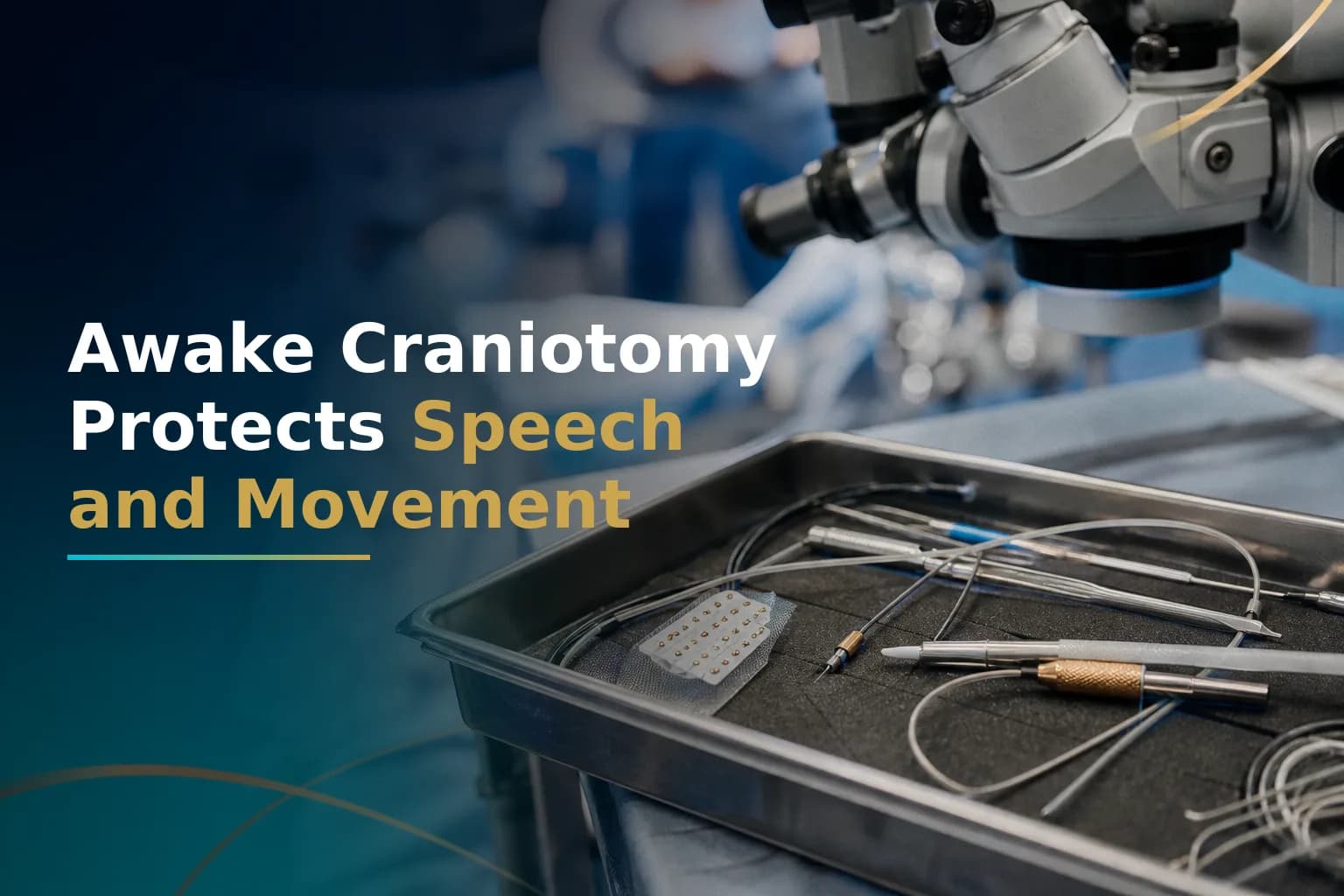
Awake Craniotomy for Glioblastoma: Speech and Motor Mapping
When a glioblastoma grows near brain areas that control speech or movement, surgeons face a tough choice: remove as much tumor as possible, or protect the functions you need for daily life. Awake craniotomy does both. You stay awake and respond during the most critical part of surgery, so your answers tell the surgeon exactly where to stop cutting.
This is not new or experimental. Major neurosurgical centers have used it for several decades, and surgeons prefer it for tumors in or near eloquent cortex - the brain regions that control language, movement, and sensation.
Learning how awake craniotomy works, who qualifies, and how it compares to standard surgery can help you ask better questions before your surgical plan is set.
What Is Awake Craniotomy?
Awake craniotomy is a neurosurgical technique in which doctors sedate you for the painful early phases - scalp incision and skull opening - then bring you to a conscious state while they map and remove tumor tissue near critical brain areas. Brain tissue has no pain receptors, so you can stay awake and talk during this phase without discomfort. Your responses guide the surgeon in real time. The goal is to remove as much tumor as safely possible while protecting speech and movement.
The NCBI StatPearls reference on awake craniotomy anesthesia explains that doctors use sedation during the more stimulating phases - skull pin placement, skin incision, craniotomy, and dural opening - and that the brain itself needs no anesthesia during the mapping phase because it lacks pain receptors.
Who Is a Candidate for Awake Craniotomy?
Doctors use this technique when the tumor sits in or close to brain regions that control language, movement, or sensation. Research from technical mapping studies at major neurosurgical programs shows that 46 to 61 percent of gliomas are located in or near eloquent brain regions. This helps explain why awake craniotomy is one of the more common advanced surgical techniques for high-grade glioma care.
Typical eligibility criteria include:
- Tumor located in or adjacent to the language cortex, motor strip, or sensory cortex
- Ability to follow instructions and communicate clearly before surgery
- No severe pre-existing aphasia (loss of ability to communicate) that already prevents reliable testing
- No significant anxiety disorder or claustrophobia that would prevent cooperation during the awake phase
- No large midline shift or severely raised intracranial pressure
- Tumor in the upper brain (supratentorial), not the cerebellum or brainstem
Some patients are excluded even if their tumor location would otherwise call for this approach. Anyone who cannot cooperate reliably during intraoperative testing is not a suitable candidate. The National Brain Tumor Society describes a pre-surgical evaluation process that confirms whether a patient can participate before a date is set.
Tumor location is one of the most important factors in planning surgery and radiation. The article on How Glioblastoma Tumor Location Shapes Surgery Options covers how proximity to critical brain structures affects the full treatment plan, not just the surgical decision.
How Does Awake Craniotomy Compare to Surgery Under General Anesthesia?
Both approaches aim for maximum safe resection. The key difference is how the surgeon finds and protects critical brain tissue during surgery. Under general anesthesia, the team uses preoperative imaging and neuronavigation systems to estimate where critical areas lie. In an awake craniotomy, the patient's real-time responses confirm this directly instead of relying on estimates.
| Comparison Axis | Awake Craniotomy | General Anesthesia Craniotomy |
|---|---|---|
| Patient state during resection | Sedated for skull opening; awake and communicating during mapping and tumor removal | Fully anesthetized throughout the entire operation |
| Best suited for | Tumors in or near language cortex, motor strip, or sensory cortex; cooperative patients | Tumors away from eloquent cortex; patients unable to cooperate or with severe pre-existing deficits |
| Extent of resection near eloquent areas | May allow more complete resection - surgeons can work closer to critical tissue safely because they get real-time feedback | Typically more conservative near eloquent cortex, because surgeons cannot assess function in real time |
| Risk of persistent neurological deficit | Low - a 2020 systematic review found persistent deficits in only 1.9 percent of awake craniotomy patients | Higher when the tumor borders eloquent cortex and surgeons cannot assess function in real time |
| Patient experience | Requires psychological preparation; most patients in published reports say the experience is less distressing than they expected | No awareness during surgery; may feel simpler before the operation, but offers less functional protection near critical areas |
Sources: Awake craniotomy for supratentorial GBM: systematic review and meta-analysis, 14 studies and 278 patients (PMC7542985); Awake craniotomy for supratentorial gliomas: why, when and how (PMC6176819).
This matters in practice: when a GBM sits close to the motor strip or language cortex, awake craniotomy may let the surgeon remove more tumor than would be safe under general anesthesia alone. Larger tumor removal is linked to longer survival in glioblastoma. The article on How Glioblastoma Extent of Resection Affects Survival explains the link between how much tumor is removed and long-term outcome.
What Happens During Awake Craniotomy - Step by Step
The procedure unfolds in distinct phases that most patients find easier to experience after preparation than they expected.
- Pre-surgical evaluation. A speech pathologist or neuropsychologist tests your language and motor function before the operation. This baseline becomes the benchmark the team compares against during surgery.
- Sedation and skull opening. The scalp is numbed with a local anesthetic block, and you receive sedative and pain relief medication. The surgeon opens the skull and the covering of the brain - called the dura. You are sedated during this phase.
- Awakening. The anesthesia team slowly reduces sedation. You are asked to count, name objects, or move your hand. The team confirms you are comfortable and alert before mapping begins.
- Cortical and subcortical mapping. The surgeon applies a small electrical stimulation probe to the brain surface. When stimulation disrupts a function - causing speech to pause momentarily, for example - that area is marked as critical and protected. This mapping shows where resection must stop.
- Tumor removal. The surgeon removes tumor tissue, pausing regularly to retest your responses. If speech slurs or a hand weakens, the surgeon adjusts direction or depth.
- Closure. Once resection is complete or a safe boundary is confirmed, you may be lightly sedated again for skull and scalp closure. Total surgery time typically ranges from four to eight hours, depending on tumor size and location.
What Are the Risks?
Awake craniotomy has a good safety record when performed at high-volume centers, but specific risks apply.
The most common complication during surgery is seizure. Published research on awake craniotomy for supratentorial gliomas reports that seizures during surgery happen in 2 to 20 percent of procedures. Most are brief and stop with cold saline irrigation of the brain surface. A small number require switching to general anesthesia, which stops functional testing but does not increase overall risk to the patient.
Early speech or motor problems after surgery are common. The 2020 systematic review in the table above found that early post-surgery neurological deficits occurred in 34.5 percent of awake craniotomy patients - but persistent deficits occurred in only 1.9 percent. This gap matters. Most deficits in the days after surgery reflect temporary swelling or tissue response, not permanent damage.
Other risks include:
- Psychological distress during the awake phase, though most patients in published case series say the experience is tolerable with good preparation
- Inability to complete the awake phase - referred to as a failed awake craniotomy - with a switch to general anesthesia as a safety backup
- Standard surgical risks: infection, bleeding, blood clots, and medication reactions
Does Awake Craniotomy Improve Survival in Glioblastoma?
The survival evidence is mixed and should be read carefully. One multicenter retrospective comparison of high-grade glioma resection found a median overall survival of 1.70 years in awake craniotomy patients compared to 1.25 years in the general anesthesia group. But the study was retrospective, and the survival difference did not hold uniformly across all analyses. Retrospective data cannot fully account for the possibility that awake craniotomy patients were selected because their tumors were in locations where greater resection was achievable - not that the technique itself added months of life.
What the data more consistently supports is this: awake craniotomy lets surgeons remove more tumor, with less risk of permanent neurological deficit, in patients whose tumors border eloquent cortex. Both outcomes affect the full treatment picture. Greater resection reduces the residual tumor burden that chemoradiation must address. And preserved neurological function means you are more likely to complete the full course of treatment - including temozolomide and radiation, and possibly Tumor Treating Fields - without delays or dose reductions that compromise effectiveness.
A patient who loses significant speech or motor function during surgery may not complete standard-of-care therapy on schedule. This is not a separate concern from survival strategy - it is part of it.
Where Should You Have This Surgery?
Experience matters more than almost any other single factor in awake craniotomy. The National Brain Tumor Society notes that some NCI-Designated Cancer Centers perform nearly 100 awake craniotomies annually, while smaller institutions may do only a handful each year. Volume reflects how familiar the surgical team is with awake-phase anesthesia protocols, the neuropsychology support that prepares patients before surgery, and the ability to manage surgery complications without losing the ability to complete the mapping.
If your planned surgical center performs fewer than 20 awake craniotomies per year, it is reasonable to ask about that volume and to consider whether a second opinion at a higher-volume center is worthwhile before committing to a surgical date. This is a procedure where the team's specific hands-on experience has a measurable effect on outcomes.
For patients researching centers outside their home country, major neurosurgical programs in India now perform awake craniotomy with intraoperative cortical mapping at substantially lower cost than comparable procedures in the US or UK. The article on Advanced Glioblastoma Surgery in India: Costs vs US and UK explains what is available and how costs compare for international patients.
If you are weighing a surgical plan and want an independent expert review before committing to a date, you can arrange a remote second opinion through Art of Healing Cancer to have the MRI, pathology report, and proposed surgical approach reviewed by a specialist without traveling.
When to Talk to Your Doctor
Raise the question of awake craniotomy if your tumor is in or near the language cortex, motor strip, or sensory cortex - and especially if you have already noticed any change in speech, word-finding, or limb strength. Ask your neurosurgeon directly: Is this tumor in eloquent cortex? How many awake craniotomies does this center perform each year? What is the plan if I cannot cooperate during the awake phase? What baseline neurological testing will happen before surgery?
If you want an independent view on whether awake craniotomy is being appropriately recommended - or appropriately omitted - a second opinion before surgery is a reasonable step. You can upload your imaging and reports at the Glioblastoma Center patient-journey page to request a remote review of your surgical plan.
This article is for general information and is not a substitute for medical advice. Always consult your oncologist or care team about your specific situation.