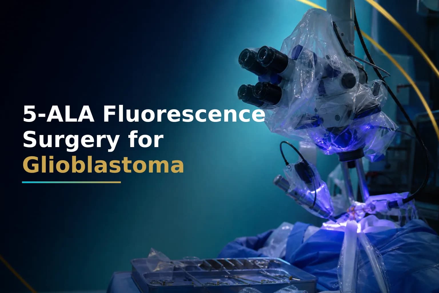
When a surgeon removes a glioblastoma, the boundary between tumor and healthy brain looks nearly identical under ordinary white light. That ambiguity makes complete removal harder than it should be. 5-ALA fluorescence-guided surgery changes this by using a drug taken a few hours before the operation to make glioblastoma cells glow a distinct pink-red under blue-violet microscope light, giving the surgical team a live visual map during the resection. This article explains how the technique works, what the clinical evidence shows, and what to ask before your surgery is scheduled.
What Is 5-ALA Fluorescence-Guided Surgery?
5-ALA fluorescence-guided surgery (FGS) is a technique where the patient takes an oral dose of 5-aminolevulinic acid (5-ALA, brand name Gleolan) three to four hours before a craniotomy for glioblastoma. Glioblastoma cells absorb 5-ALA and convert it into a molecule called protoporphyrin IX (PpIX), which glows bright pink-red under the blue-violet light fitted to the operating microscope. Healthy brain tissue does not fluoresce in the same way. The surgeon sees a live contrast view in real time and uses it to guide how much tumor to remove and where to stop.
Why Does Extent of Resection Matter in Glioblastoma?
Removing as much tumor as safely possible - what surgeons call maximal safe resection - is one of the first decisions that shapes everything that follows in glioblastoma care. A larger resection reduces the number of tumor cells that subsequent radiation and chemotherapy must control. It also supplies more tissue for the molecular analysis - MGMT methylation status, IDH mutation testing, EGFR amplification - that shapes every treatment decision down the line. More tissue from a larger resection means more complete and reliable molecular results.
A 2022 systematic review in the Journal of Neuro-Oncology found that 5-ALA fluorescence guidance was associated with greater extent of resection and better progression-free and overall survival outcomes in high-grade glioma compared to conventional white-light surgery [1]. To understand how the volume of tumor removed connects to long-term outcomes, see our article on how glioblastoma extent of resection affects survival.
How Does 5-ALA Work Inside Tumor Cells?
5-ALA is a naturally occurring amino acid the body uses to produce heme - the compound in red blood cells. When taken in a concentrated oral dose before surgery, glioblastoma cells absorb it more aggressively than normal brain cells do. Inside the tumor cell, 5-ALA is converted step by step to protoporphyrin IX (PpIX). An enzyme that would ordinarily break PpIX down further is less active in tumor cells, so PpIX accumulates there rather than being cleared.
When the surgeon switches the operating microscope from white light to blue-violet light, the accumulated PpIX emits a bright pink-red fluorescence. Normal brain around the tumor glows faintly or not at all. The surgeon can toggle between the two light modes throughout the operation - using white light to assess anatomy, switching to blue-violet to check where tumor tissue remains before closing. That back-and-forth is what gives fluorescence guidance its practical advantage over relying on a pre-operative MRI image alone.
What the Landmark Clinical Trial Found
The evidence base for 5-ALA FGS rests largely on a Phase III randomized controlled trial by Stummer and colleagues published in Lancet Oncology. The trial enrolled 322 patients with newly diagnosed high-grade glioma and compared fluorescence-guided surgery directly to standard white-light resection. Complete removal of contrast-enhancing tumor was achieved in 65% of patients in the 5-ALA group versus 36% in the white-light group. The 6-month progression-free survival rate was 41% for 5-ALA versus 21% for white light, with no significant increase in neurological deficits [2]. On the strength of that evidence, the U.S. Food and Drug Administration approved Gleolan in June 2017 for adult patients with suspected WHO grade III or IV gliomas.
How Does 5-ALA Surgery Compare to Standard White-Light Resection?
| Factor | 5-ALA Fluorescence Surgery | Standard White-Light Surgery |
|---|---|---|
| How the surgeon sees the tumor | Tumor cells glow pink-red under blue-violet microscope light in real time | Surgeon relies on visual texture and pre-operative MRI |
| Complete resection rate | 65% of patients | 36% of patients |
| 6-month progression-free survival | 41% | 21% |
| Drug required before surgery | Oral 5-ALA (Gleolan), taken 3-4 hours before the operation | None |
| Photosensitivity risk after surgery | Yes - avoid bright light for 24-48 hours post-op | No |
| Regulatory status (US) | FDA approved June 2017 for suspected WHO grade III-IV gliomas | Standard of care globally |
Complete resection and progression-free survival figures from: Stummer et al., Lancet Oncology, 2006 - Phase III randomized controlled trial, 322 patients with newly diagnosed high-grade glioma.
The jump from 36% to 65% in complete resection rate is not a marginal improvement. It means nearly twice as many patients had all visible contrast-enhancing tumor removed when fluorescence guidance was used. The 6-month progression-free survival data tracks closely with that resection gap - suggesting that removing more tumor at surgery gives subsequent treatments a better starting position.
Does More Resection Translate to Longer Survival?
Multiple studies since the original trial have confirmed the association between 5-ALA guidance and improved survival in glioblastoma. A comparative cohort study of 343 glioblastoma patients published in Neuro-Oncology Advances found that patients who underwent 5-ALA-guided resection had improved overall survival compared to those who had white-light surgery [3]. A more recent comparative analysis confirmed that the advantage in complete resection rate was associated with better overall survival outcomes in real-world patient populations [4].
Context matters here. Glioblastoma is an infiltrating tumor - microscopic cells spread beyond what any imaging or fluorescence signal can detect. Achieving a complete resection of the contrast-enhancing tumor mass on MRI is not the same as removing every tumor cell in the brain. Surgery is the first step in a multimodal plan that also includes radiation, temozolomide chemotherapy, and in some cases Tumor Treating Fields. What the 5-ALA data consistently shows is that starting that combined treatment with a smaller residual tumor burden gives the subsequent phases better conditions to work in.
Who Is a Candidate for 5-ALA Fluorescence Surgery?
5-ALA FGS is generally used for adults with newly diagnosed or recurrent high-grade glioma where the tumor enhances on contrast-enhanced MRI and is in a location where resection is anatomically feasible. Glioblastoma is almost always IDH-wildtype, and IDH-wildtype tumors tend to accumulate strong PpIX fluorescence - making this setting the one where the data is most robust and the visual signal most reliable.
Tumors near eloquent areas - speech cortex, motor pathways - can still be resected using 5-ALA guidance, typically alongside functional brain monitoring or awake craniotomy techniques that let the surgical team protect critical function while removing tumor tissue. Our article on awake craniotomy for glioblastoma and brain mapping explains how those two approaches work together and what to expect from each.
Not every tumor fluoresces strongly. IDH-mutant lower-grade tumors tend to accumulate less PpIX, and the fluorescent signal may be weak or absent. Deep-seated glioblastomas in the brainstem or basal ganglia are more technically challenging locations, and the supporting evidence for 5-ALA in those anatomical regions is less conclusive than for cortical and subcortical tumors [1]. A neurosurgeon with experience in fluorescence-guided surgery can advise whether the signal is likely to be useful for your specific tumor location.
What Are the Side Effects and Risks?
The side effects directly related to 5-ALA itself are mild and generally short-lived. The most important is photosensitivity: skin and eyes become sensitive to bright light for up to 48 hours after taking the drug. Patients need to avoid direct sunlight and intense indoor lighting during that window. Brief unprotected exposure can produce a reaction similar to sunburn. Other reported effects include nausea, a brief drop in blood pressure, and a mild temporary rise in liver enzyme levels [5]. These effects typically resolve without specific treatment.
5-ALA does not carry the toxicity profile of chemotherapy agents. The surgical risks - bleeding, infection, and the possibility of a neurological deficit - are the same as for any craniotomy, and 5-ALA itself does not add meaningful operative risk. What it does is help the surgeon distinguish tumor from surrounding brain in real time, reducing the likelihood of leaving large amounts of tumor behind. That guidance does not replace functional monitoring; in tumors near critical brain areas, both are needed together.
Is 5-ALA Surgery Available at All Centers?
5-ALA surgery requires an operating microscope fitted with blue-violet excitation filters, the corresponding camera and display system, and surgical teams trained in interpreting the fluorescence signal intraoperatively. At major academic medical centers and dedicated brain tumor programs in the United States, Western Europe, and parts of Asia - including several large neurosurgical centers in India - 5-ALA has become a standard part of glioblastoma surgery. Availability is less consistent at community hospitals and in regions where either the modified microscope or the drug itself is not yet in place.
If your neurosurgeon has not mentioned fluorescence guidance as part of the operative plan, it is a straightforward question to raise before the operation is confirmed. If you are uncertain whether your center uses the technique routinely, or if you want an independent assessment of the surgical plan before committing, you can arrange a remote second opinion through Art of Healing Cancer. For patients weighing surgical options and costs across different countries, our article on advanced glioblastoma surgery in India and how costs compare to the US and UK covers what advanced surgical infrastructure looks like at major Indian centers and what to expect.
When to Talk to Your Doctor
Before surgery is scheduled, ask your neurosurgeon whether 5-ALA fluorescence guidance is part of the planned approach and how many glioblastoma resections the team performs with it each year. Ask what intraoperative monitoring will be used - particularly if the tumor is near speech or motor regions. If you want a second opinion on the surgical strategy or the overall treatment plan, you can upload your MRI scans and pathology reports at glioblastoma.center/patient-journey for a remote review by a specialist team.
This article is for general information and is not a substitute for medical advice. Always consult your oncologist or care team about your specific situation.